Copyright
©The Author(s) 2020.
World J Clin Cases. May 26, 2020; 8(10): 1944-1949
Published online May 26, 2020. doi: 10.12998/wjcc.v8.i10.1944
Published online May 26, 2020. doi: 10.12998/wjcc.v8.i10.1944
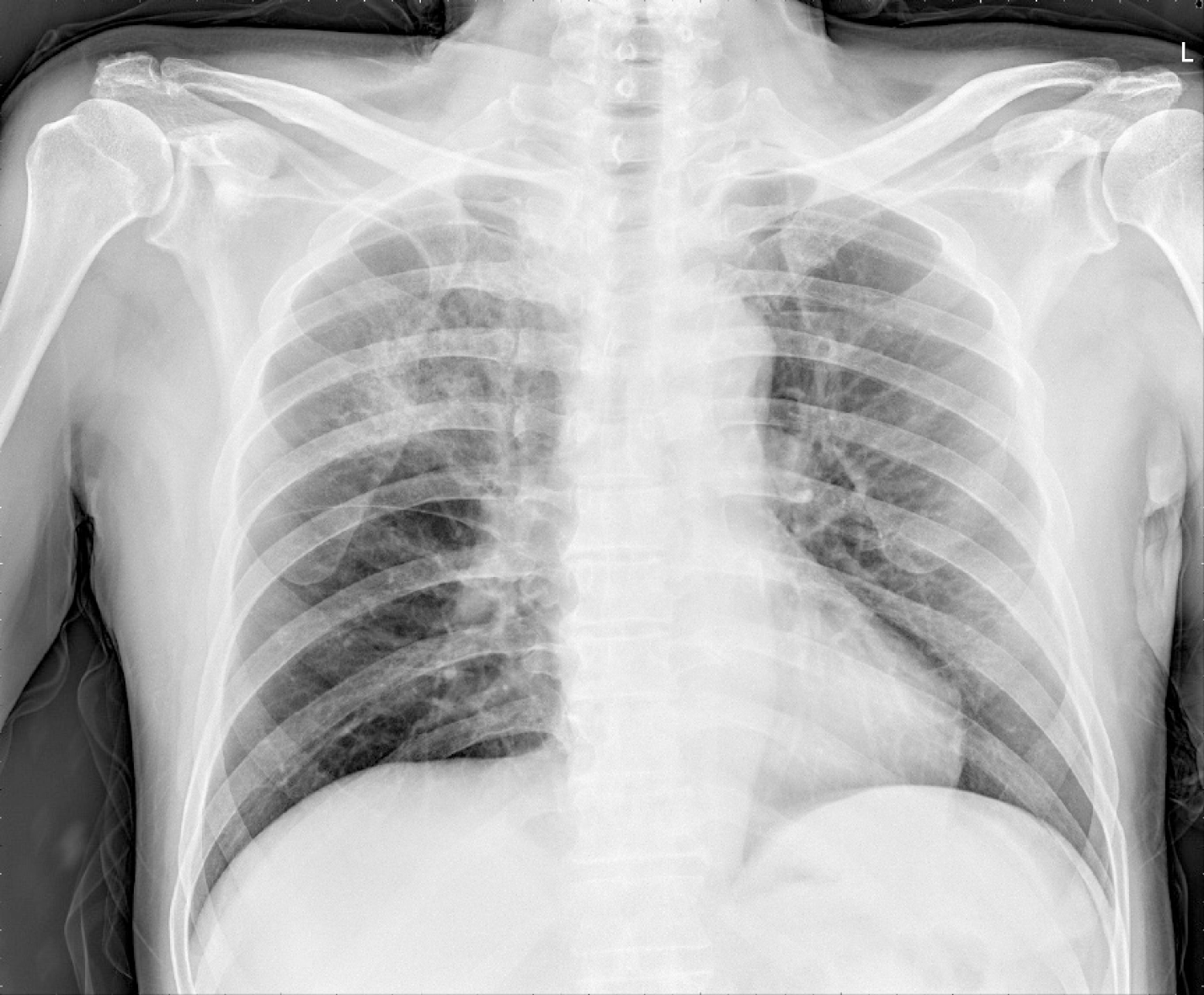
Figure 1 Chest radiography.
Consolidation is seen in the right upper lobe of the lung.
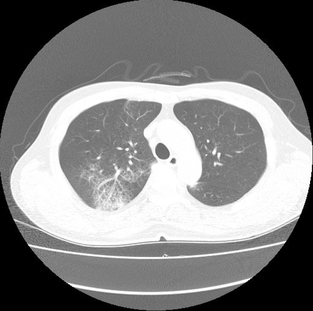
Figure 2 Contrast-enhanced chest computed tomography (Axial plane).
Chest computed tomography showing patchy ground-glass opacity in the right upper lobe.
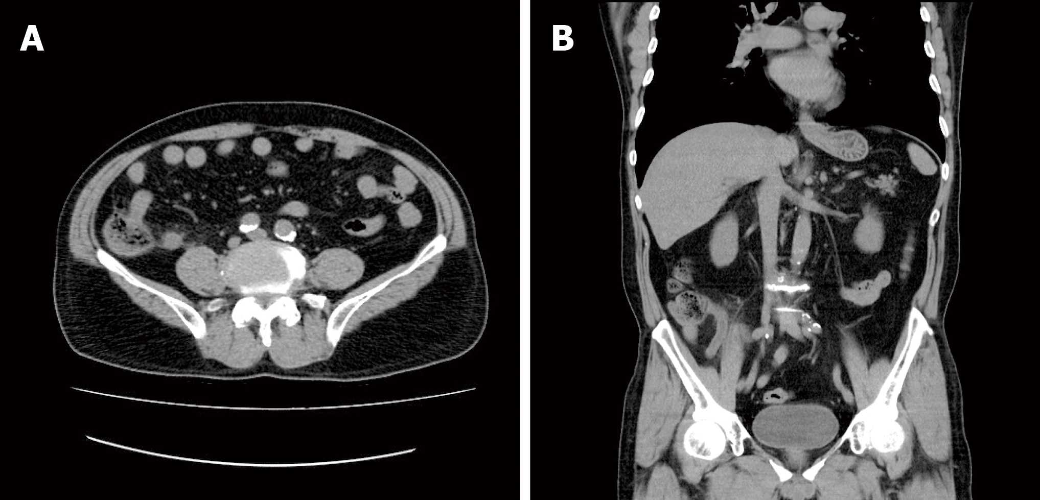
Figure 3 Non-contrast-enhanced abdomen computed tomography.
A: Axial plane; B: Coronal plane. Abdomen computed tomography showing an 11-mm dilatation of the appendix.
- Citation: Kim C, Kim JK, Yeo IH, Choe JY, Lee JE, Kang SJ, Park CS, Kwon KT, Hwang S. Appendectomy in patient with suspected COVID-19 with negative COVID-19 results: A case report. World J Clin Cases 2020; 8(10): 1944-1949
- URL: https://www.wjgnet.com/2307-8960/full/v8/i10/1944.htm
- DOI: https://dx.doi.org/10.12998/wjcc.v8.i10.1944


