Copyright
©The Author(s) 2020.
World J Clin Cases. May 26, 2020; 8(10): 1832-1847
Published online May 26, 2020. doi: 10.12998/wjcc.v8.i10.1832
Published online May 26, 2020. doi: 10.12998/wjcc.v8.i10.1832
Figure 1 Schematic illustration of traditional classifications and representative modifications of the keystone design perforator island flap.
A: Type I keystone design perforator island flap (KDPIF) (skin incision only); B: Type IIA KDPIF (division of the deep fascia along the outer curvilinear line); C: Type IIB KDPIF (division of the deep fascia and skin graft to the secondary defect); D: Type III KDPIF (opposing keystone flaps designed to create a double-keystone flap); E: Type IV KDPIF (keystone flap with undermining of up to 50% of the flap subfascially); F: The Ω-variant KDPIF (defect closure in the fish-mouth fashion). Further undermining of the flap (blue-colored circle) with preserving the central hot spot of perforators (red-colored x marks); G: Sydney melanoma unit modification (maintenance of a skin bridge along the greater arc of the KDPIF). KDPIF: Keystone design perforator island flap; SMU: Sydney melanoma unit.
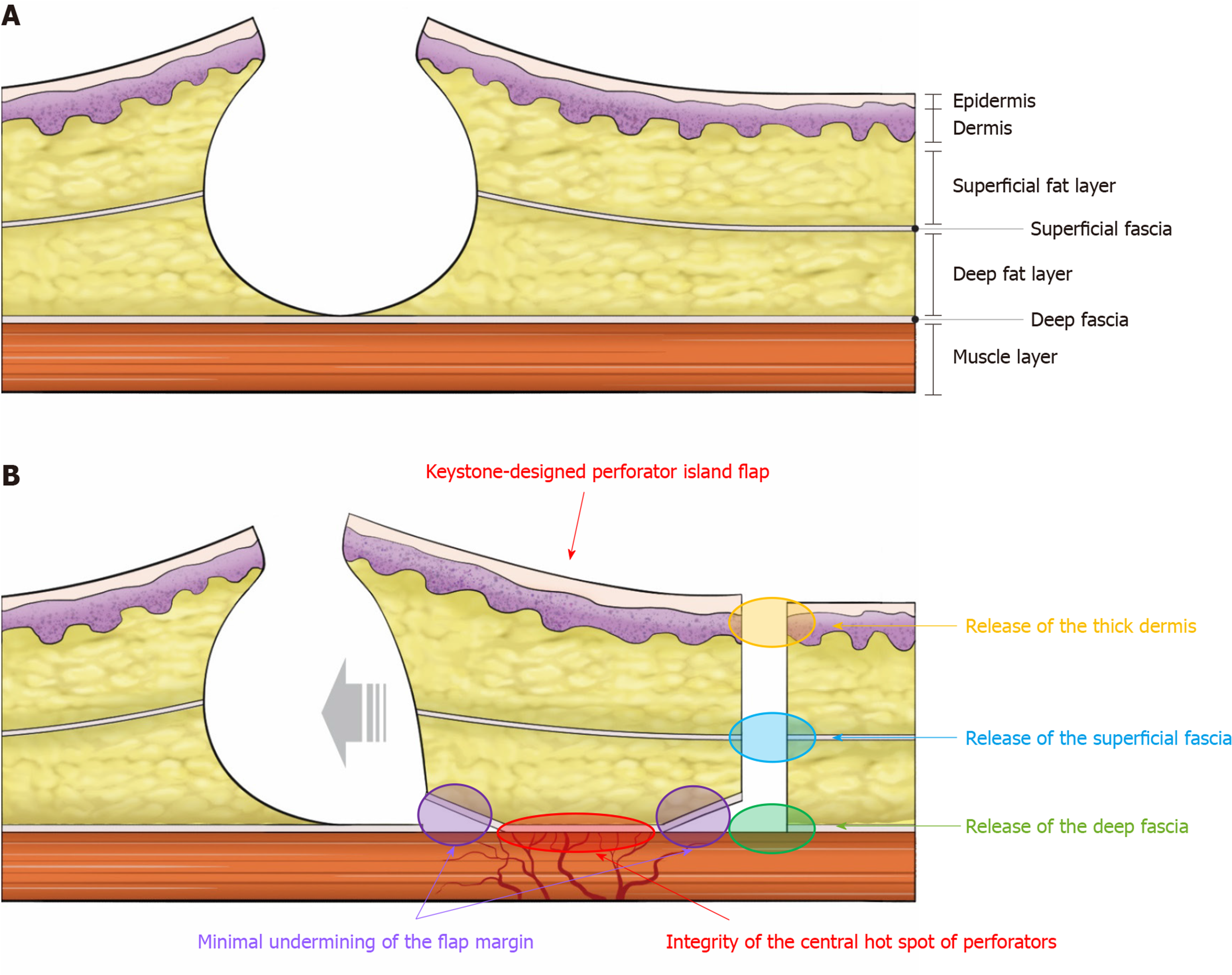
Figure 2 Schematic diagram of the keystone design perforator island flap movement.
A: Cross-sectional diagram; B: The keystone design perforator island flap movement on the back is provoked by three main factors. The first factor (orange-colored oval) is the release of the thick dermis at the back; the second (blue-colored oval) is the release of the superficial fascia; and the last (green-colored oval) is the release of the deep fascia. Then, minimal flap undermining of the flap margin (purple-colored circle) is performed to preserve the integrity of the “central hot spot” of the perforators (red-colored oval). (Reprinted from Yoon et al[14], with permission from Springer Nature).
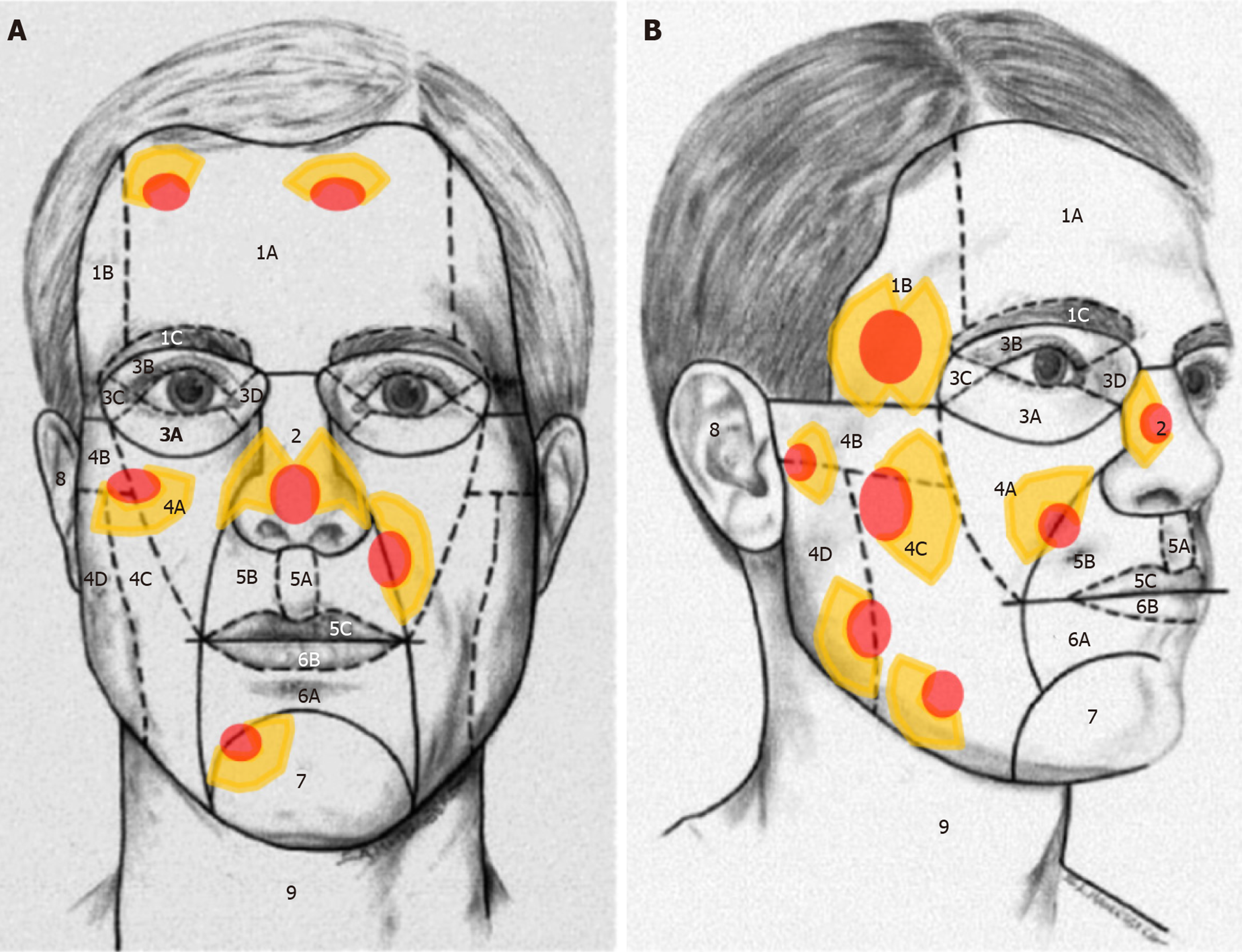
Figure 3 Schematic diagram showing the relaxed skin tension line-oriented keystone design perforator island flap considering the facial aesthetic unit concept.
Red-colored ellipses represent defects and yellow-colored figures represent the design of the keystone design perforator island flap. Frontal (A) and profile views (B) of the aesthetic units and subunits of the face. 1: Forehead unit (1A, central subunit; 1B, lateral subunit; 1C, eyebrow subunit); 2: Nasal unit; 3: Eyelid units (3A, lower-lid unit; 3B, upper-lid unit; 3C, lateral canthal subunit; 3D, medial canthal subunit); 4: Cheek unit (4A, medial subunit; 4B, zygomatic subunit; 4C, lateral subunit; 4D, buccal subunit); 5: Upper-lip unit (5A, philtrum subunit; 5B, lateral subunit; 5C, mucosal subunit); 6: Lower-lip unit (6A, central subunit; 6B, mucosal subunit); 7: Mental unit; 8: Auricular unit; 9: Neck unit. (Reprinted from Yoon et al[1], with permission from Wolters Kluwer, which was originally reprinted from Fattahi, with permission from Elsevier).
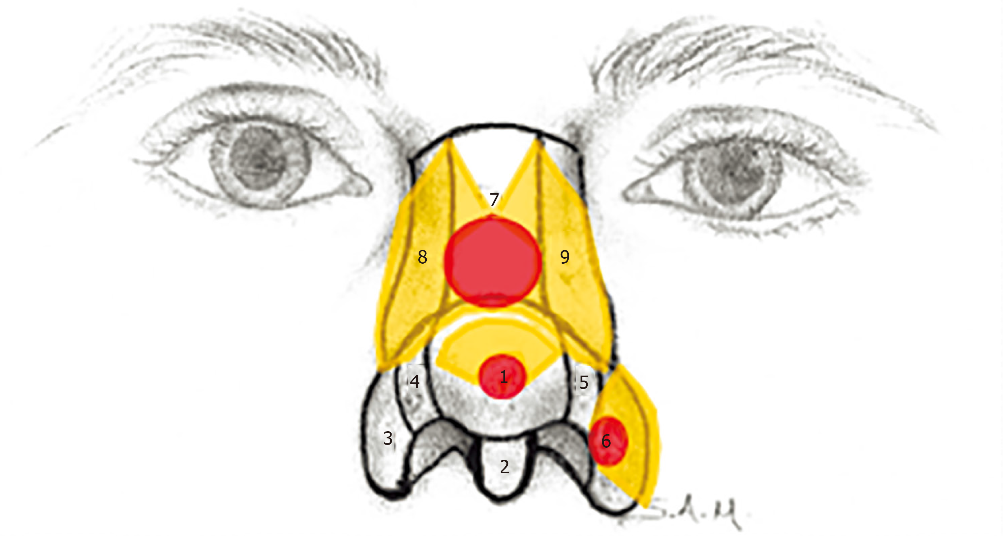
Figure 4 Schematic diagram showing the relaxed skin tension line-oriented keystone design perforator island flap considering the nasal unit.
Red-colored ellipses represent defects and yellow-colored figures represent the design of keystone design perforator island flap. 1: Tip subunit; 2: Columellar subunit; 3, 6: Right and left alar base subunits; 4, 5: Right and left alar side wall subunits; 7: Dorsal subunit; 8, 9: Right and left dorsal side wall subunits. (Reprinted from Yoon et al[1], with permission from Wolters Kluwer, which was originally reprinted from Fattahi, with permission from Elsevier).
Figure 5 A 67-year-old woman was diagnosed with basal cell carcinoma on the nose after a punch biopsy.
There were two lesions in the right dorsal subunit and the left alar side wall subunit of the nasal unit. A: She underwent wide excision of each lesion with a 3-mm safety margin, and each final defect was measured to be 1.8 cm x 1.8 cm on the right side and 1.5 cm x 1.5 cm on the left side; B, C: We covered defects with each Ω-variant Type IIA keystone design perforator island flap (flap size: 3.5 cm × 5 cm) on the right side and a Ω-variant Type IIA keystone design perforator island flap (flap size: 2 cm × 3.5 cm) on the left side. Final suture lines were located within and along each facial subunit; D: Postoperative clinical photograph after 12 mo.
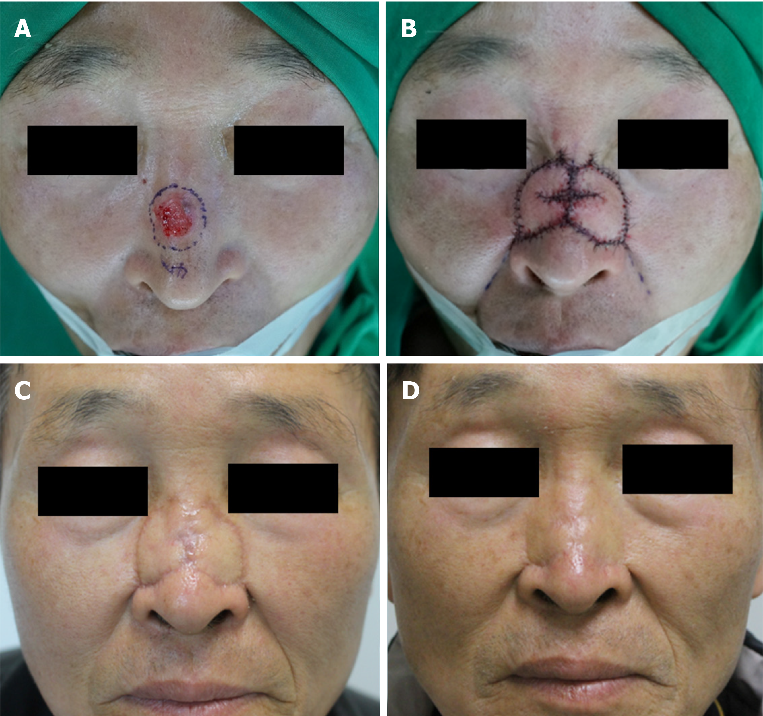
Figure 6 A 62-year-old man was diagnosed with basal cell carcinoma on the nose (dorsal subunit of the nasal unit) after a punch biopsy.
A: He underwent wide excision with a 4-mm safety margin, and the final defect was measured to be 3 cm × 2.5 cm; B: We covered the defect with bilateral Ω-variant keystone-designed perforator island flaps (each flap size was 1.5 cm × 3.5 cm) from both dorsal side wall subunits; C: Postoperative clinical photograph after 1 mo of follow-up; D: Postoperative clinical photograph after 12 mo of follow-up. (Reprinted from Yoon et al[1], with permission from Wolters Kluwer).
Figure 7 An 82-year-old man was diagnosed with basal cell carcinoma on the right ala after a punch biopsy.
A: The lesion was located on the right alar base subunit of the nasal unit; B: He underwent wide excision of the lesion with a 3-mm safety margin, the final defect was measured to be 2.5 cm × 2.8 cm; C: We covered the defect with a Type IIA keystone design perforator island flap (flap size: 4 cm × 6 cm) from the medial subunit of the right cheek unit. Final suture lines were located within and along each facial subunit; D: Postoperative clinical photograph after 3 mo.
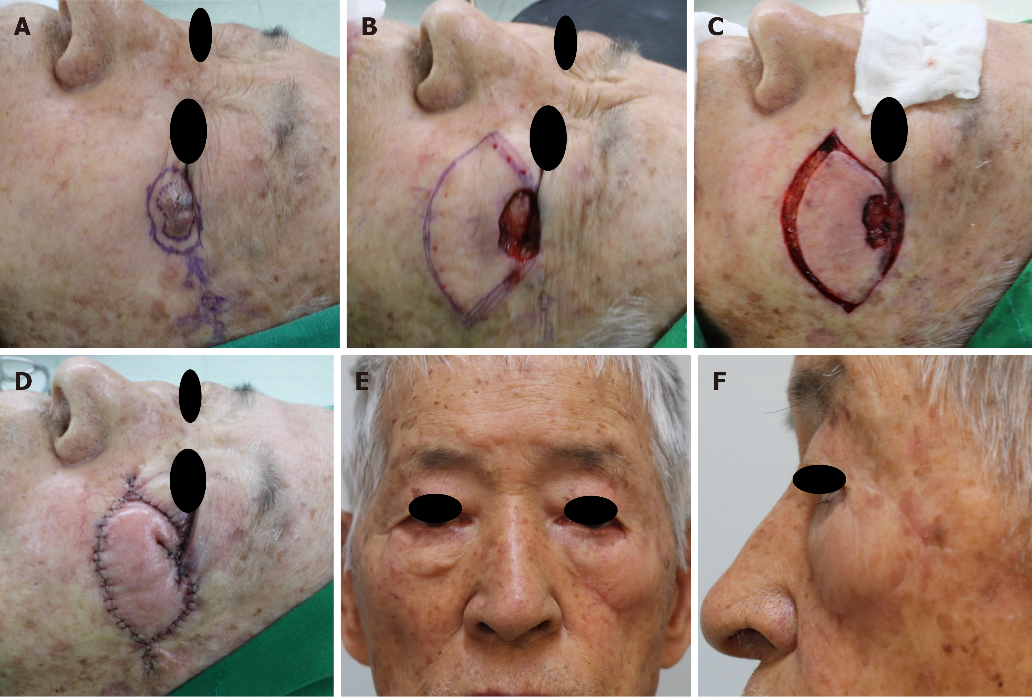
Figure 8 A 65-year-old man was diagnosed with basal cell carcinoma on the right medial canthal area after a punch biopsy.
A: The lesion was located on the medial canthal subunit and lower-lid subunit of the right eyelid unit; B: He underwent wide excision of the lesion with a 4-mm safety margin, the final defect was measured to be 1.8 cm × 2.5 cm; C, D: We covered the defect with a Ω-variant Type IIA keystone design perforator island flap (flap size: 2.5 cm × 6 cm) from the right dorsal subunit and dorsal side wall subunit of the nasal unit. Final suture lines were located within and along each facial subunit; E, F: Postoperative clinical photograph after 6 mo.
Figure 9 An 86-year-old man was diagnosed with basal cell carcinoma on the left lateral canthal area after a punch biopsy.
A: The lesion was located on the lateral canthal subunit and lower-lid subunit of the left eyelid unit; B: He underwent wide excision of the lesion with a 3-mm safety margin, the final defect was measured to be 2.5 cm × 3 cm; C, D: We covered the defect with a Ω-variant Type IIA keystone design perforator island flap (flap size: 4 cm × 8.5 cm) from the zygomatic subunit and medial subunit of the left cheek unit. Final suture lines were located within and along each facial subunit, and parallel to facial relaxed skin tension lines; E, F: Postoperative clinical photograph after 12 mo.
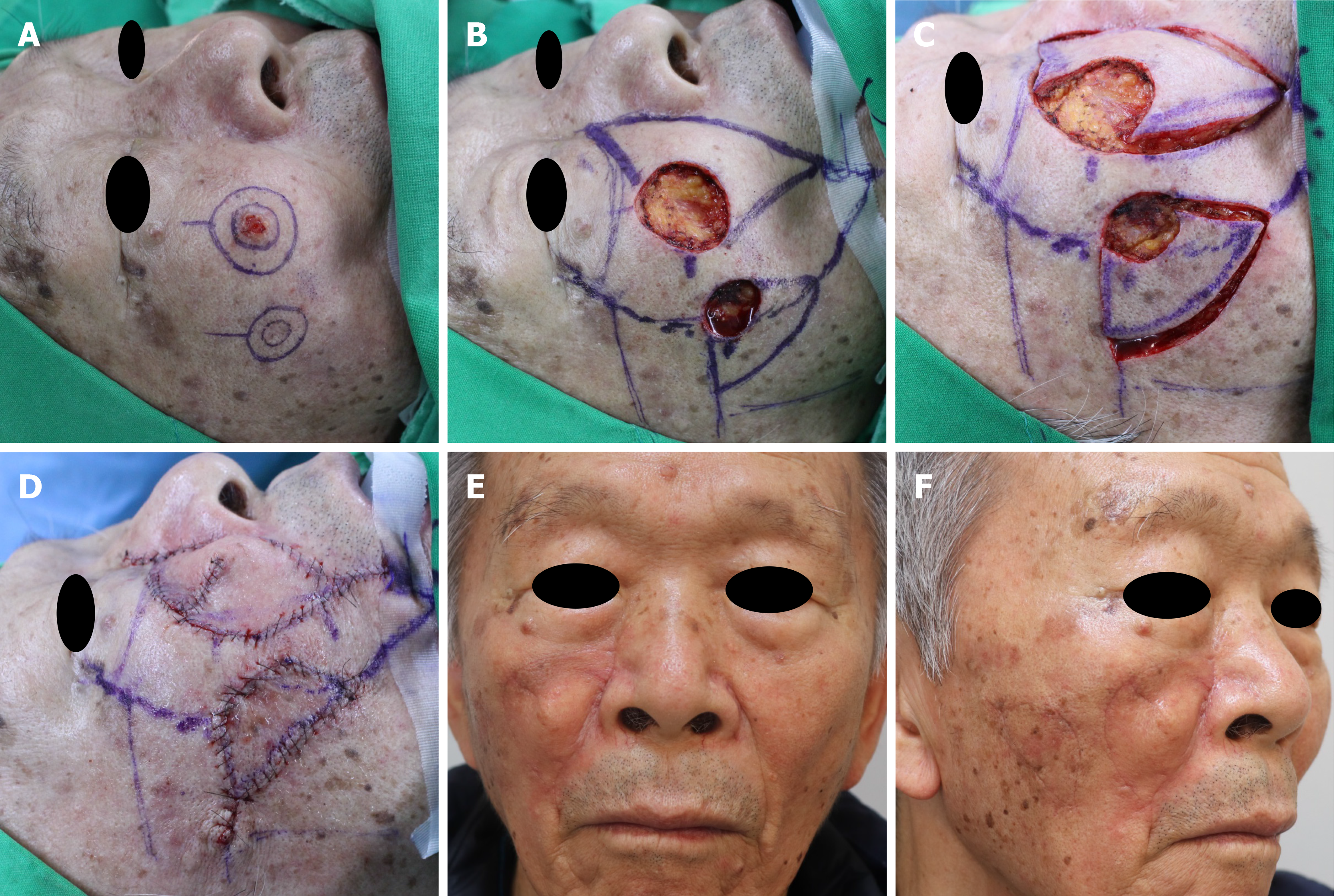
Figure 10 A 77-year-old man was diagnosed with basal cell carcinoma on the right cheek after a punch biopsy.
A: There were two lesions in the medial subunit and the lateral subunit of the right cheek unit; B: He underwent wide excision of each lesion with a 5-mm safety margin, and each final defect was measured to be 2.6 cm × 2.6 cm in the medial subunit and 1.5 cm × 1.5 cm in the lateral subunit; C, D: We covered the medial subunit defect and the lateral subunit defect with a Ω-variant Type IIA keystone design perforator island flap (flap size: 3.5 cm × 7 cm) and a Type I keystone design perforator island flap (flap size: 2 cm × 4 cm) in consideration of facial relaxed-skin tension lines and the facial subunit concept, respectively. Final suture lines were located within and along each facial subunit; E, F: Postoperative clinical photographs after 3 mo.
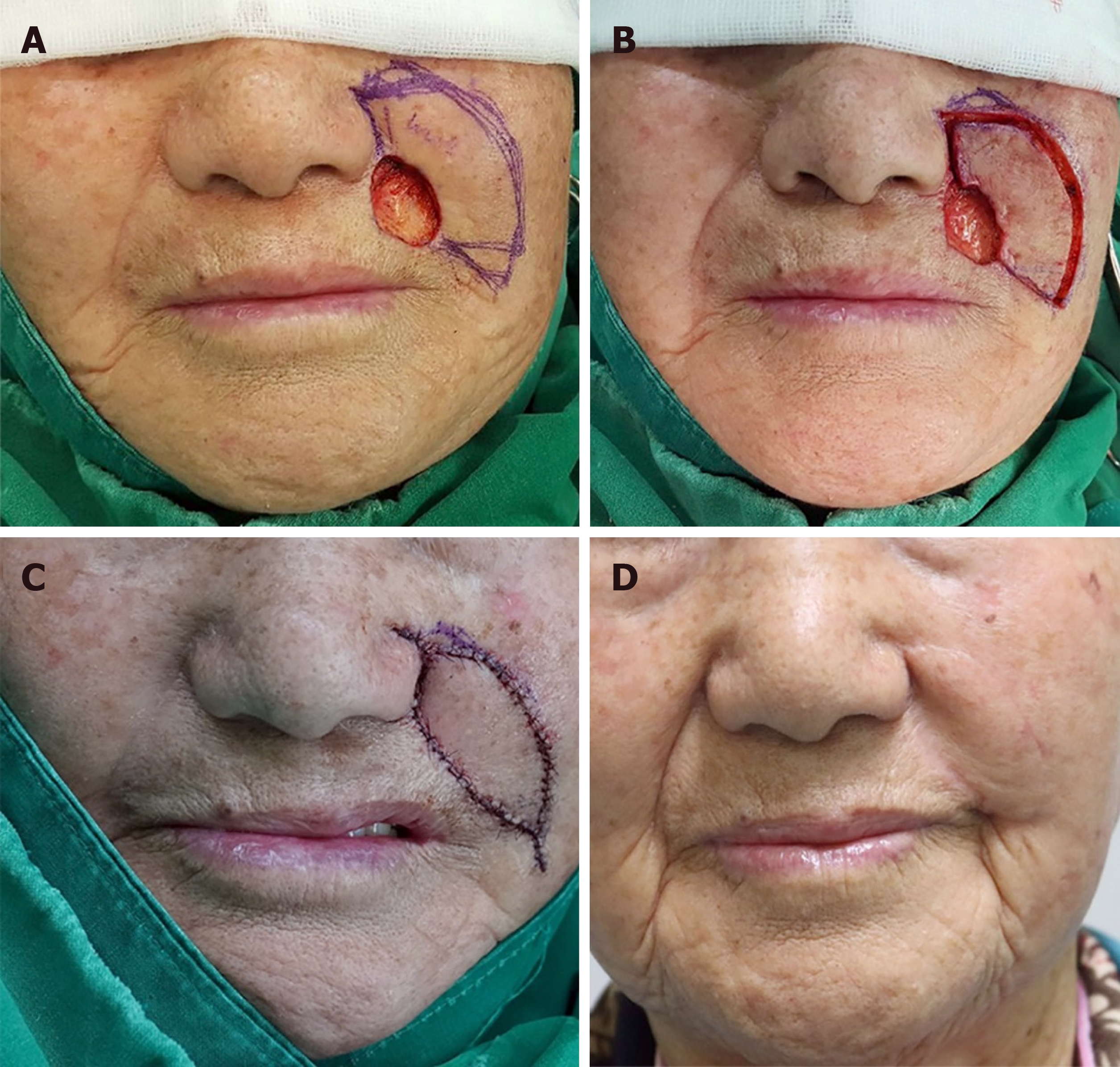
Figure 11 An 82-year-old woman was diagnosed with basal cell carcinoma in the left nasolabial fold area (medial subunit of the cheek unit) by punch biopsy.
A: She underwent a wide excision with a 4-mm safety margin and the final defect was measured to be 2 cm × 3 cm; B, C: We covered the defect with a Type IIA keystone-designed perforator island flap (flap size: 2.5 cm × 5.5 cm) from the upper-lateral side of the defect; D: Postoperative clinical photograph after 6 mo of follow-up. (Reprinted from Yoon et al[1], with permission from Wolters Kluwer).
Figure 12 A 63-year-old woman was diagnosed with basal cell carcinoma on the right forehead after a punch biopsy.
A: The lesion was located on the right lateral subunit of the forehead unit; B: She underwent wide excision of the lesion with a 4-mm safety margin, the final defect was measured to be 1.5 cm × 1.5 cm; C: We covered the defect with a Type IIA keystone design perforator island flap (flap size: 2 cm × 4.5 cm). Final suture lines were located on the periphery of forehead unit, and parallel to forehead relaxed skin tension lines and hairlines; D: Postoperative clinical photograph after 6 mo.
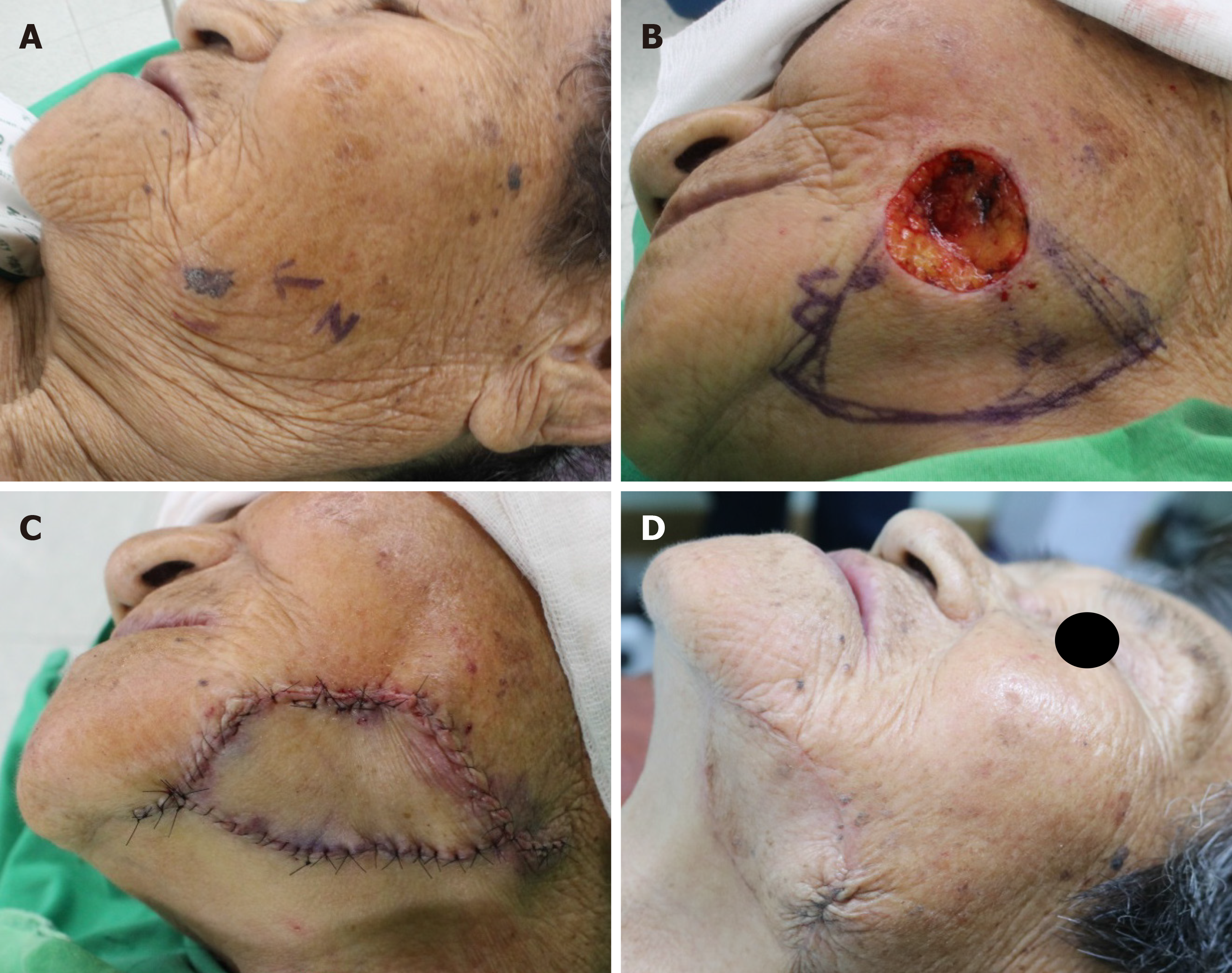
Figure 13 An 82-year-old woman was diagnosed with basal cell carcinoma on the left mandible border after a punch biopsy.
A: The lesion was located on the lower part of the left lateral subunit of the cheek unit; B: She underwent wide excision of the lesion with a 5-mm safety margin, the final defect was measured to be 3 cm × 3 cm; C: We covered the defect with a Type IIA keystone design perforator island flap (flap size: 3.5 cm × 8 cm) in the lateral subunit of the cheek unit. Final suture lines were located within the lowest part of the lateral subunit of the cheek unit; D: Postoperative clinical photograph after 4 mo.
- Citation: Lim SY, Yoon CS, Lee HG, Kim KN. Keystone design perforator island flap in facial defect reconstruction. World J Clin Cases 2020; 8(10): 1832-1847
- URL: https://www.wjgnet.com/2307-8960/full/v8/i10/1832.htm
- DOI: https://dx.doi.org/10.12998/wjcc.v8.i10.1832


