Copyright
©The Author(s) 2020.
World J Clin Cases. May 26, 2020; 8(10): 1756-1762
Published online May 26, 2020. doi: 10.12998/wjcc.v8.i10.1756
Published online May 26, 2020. doi: 10.12998/wjcc.v8.i10.1756
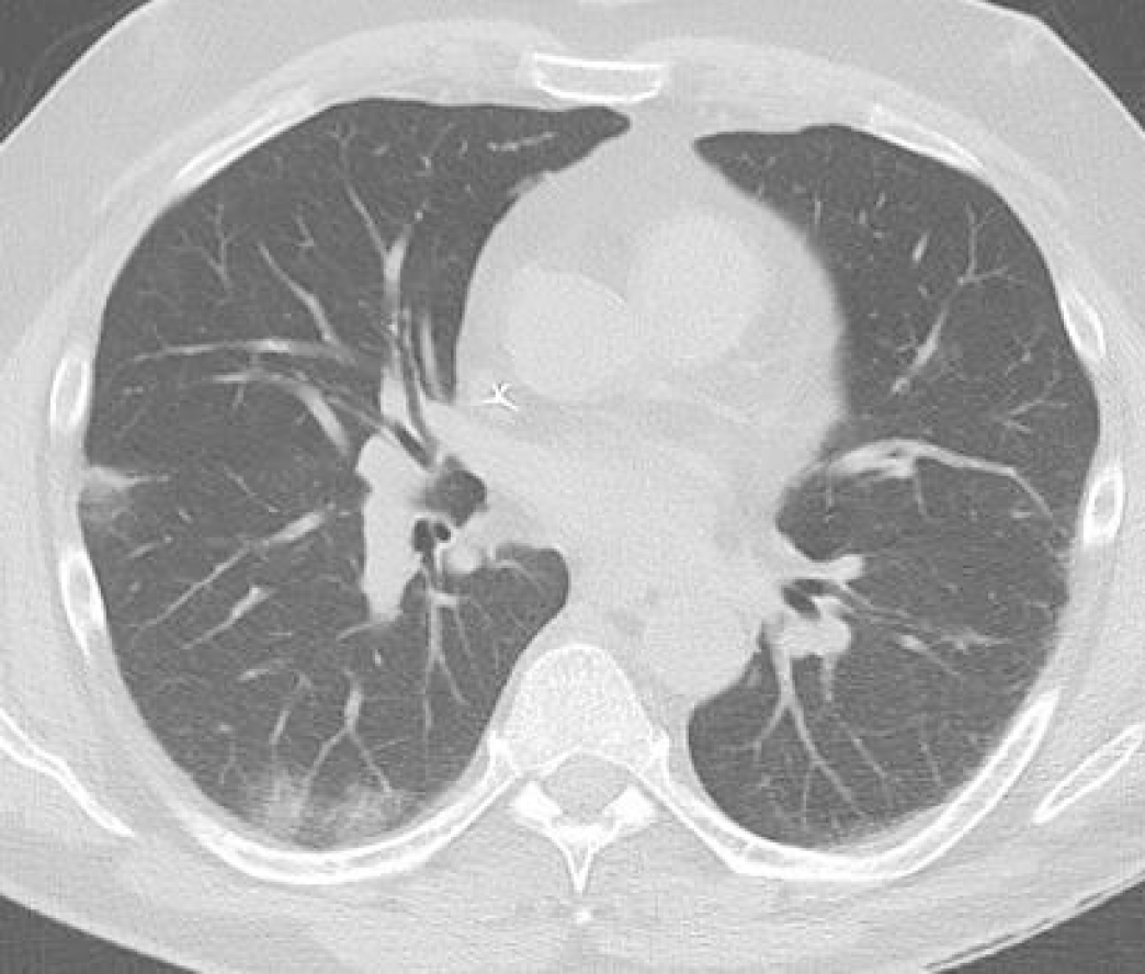
Figure 1 Chest computed tomography-scan revealing coronavirus disease 2019 typical thoracic signs of peripheral ground glass opacities.
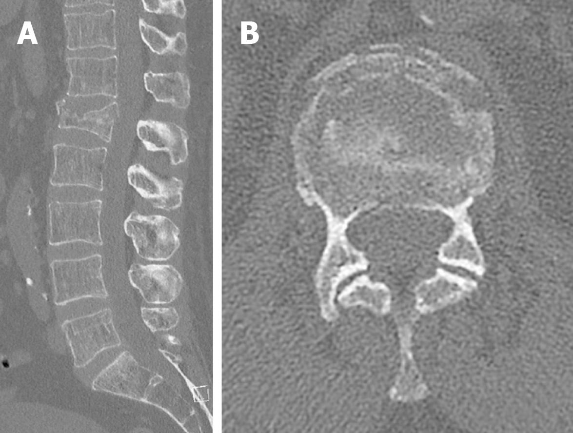
Figure 2 Spine computed tomography scan.
A: Sagittal view; B: Axial view. The images showed that the stable vertebral compression L1 fracture without significant posterior wall involvement. A conservative treatment was carried out.
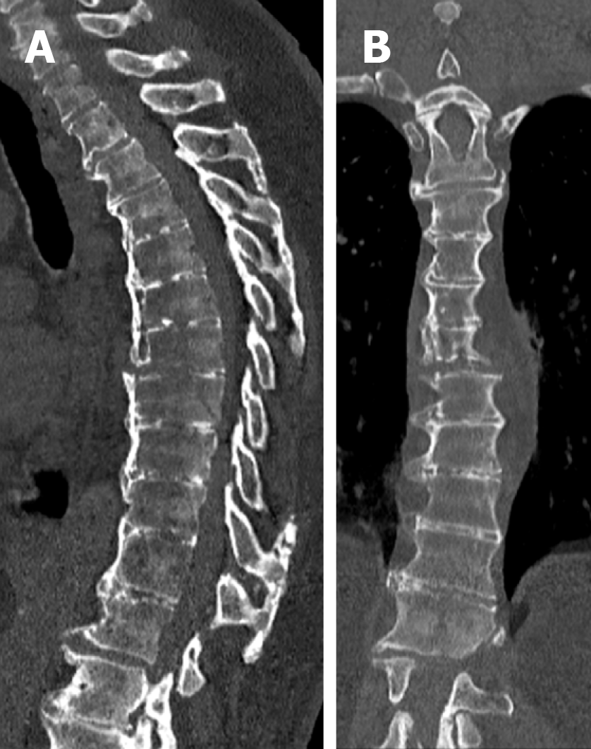
Figure 3 Sagittal and coronal spine computed tomography scan showing an unstable non-neurologic T7-T8 fracture on ankylosing spine.
A: Sagittal; B: Coronal.
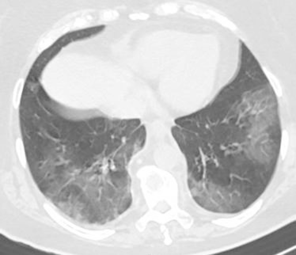
Figure 4 Coronavirus disease 2019 typical thoracic computed tomography-scan with bilateral ground glass opacities.
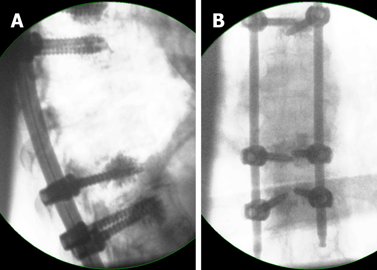
Figure 5 Intra-operative lateral and anteroposterior fluoroscopic control after T5-T10 cement augmented percutaneous posterior fixation.
A: Intra-operative lateral; B: Anteroposterior.
- Citation: Prost S, Charles YP, Allain J, Barat JL, d'Astorg H, Delhaye M, Eap C, Zairi F, Guigui P, Ilharreborde B, Meyblum J, Le Huec JC, Lonjon N, Lot G, Hamel O, Riouallon G, Litrico S, Tropiano P, Blondel B, the French Spine Surgery Society. French Spine Surgery Society guidelines for management of spinal surgeries during COVID-19 pandemic. World J Clin Cases 2020; 8(10): 1756-1762
- URL: https://www.wjgnet.com/2307-8960/full/v8/i10/1756.htm
- DOI: https://dx.doi.org/10.12998/wjcc.v8.i10.1756


