Copyright
©The Author(s) 2020.
World J Clin Cases. Jan 6, 2020; 8(1): 97-102
Published online Jan 6, 2020. doi: 10.12998/wjcc.v8.i1.97
Published online Jan 6, 2020. doi: 10.12998/wjcc.v8.i1.97
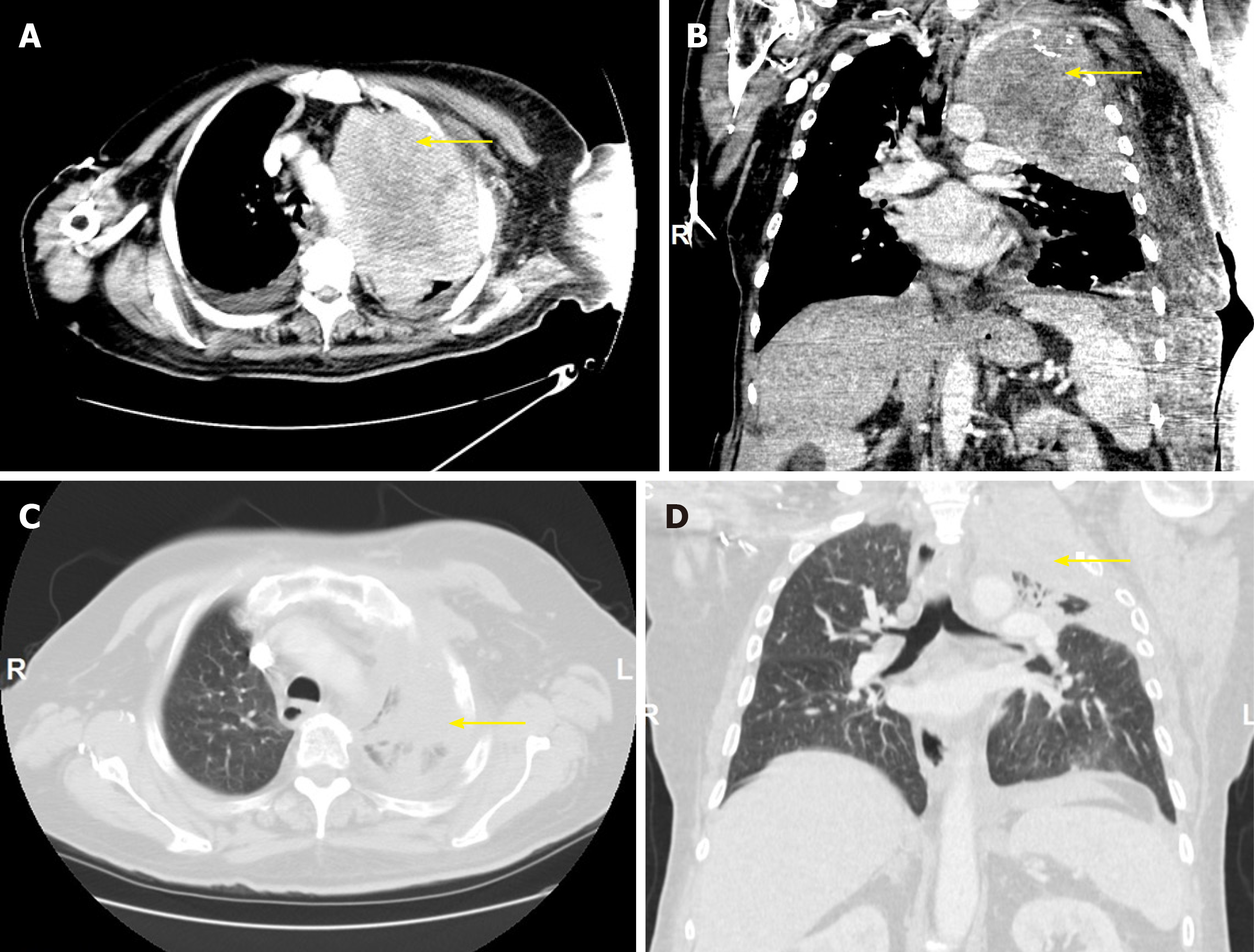
Figure 1 Chest computed tomography.
A, B: Prior to initiation of pembrolizumab, with yellow arrows pointing to a left upper lobe primary measuring 14.1 cm × 10.6 cm × 14.9 cm. C, D: After 5 cycles of pembrolizumab, with yellow arrows pointing to a markedly decreased left upper lobe mass measuring 8.0 cm × 7.7 cm × 6.8 cm.
Figure 2 Histological lung section.
A: Hematoxylin and eosin staining showing sarcomatoid carcinoma, with visible spindle cells and rare epithelioid cells (magnification, 100 ×); B: Immunohistochemical staining showing sarcomatoid carcinoma with neoplastic cells positive for CK7 (magnification, 200 ×); C: Hematoxylin and eosin staining showing sarcomatoid carcinoma, with cells positive for programmed death-ligand-1 and tumor proportion score greater than 50% (magnification, 400×).
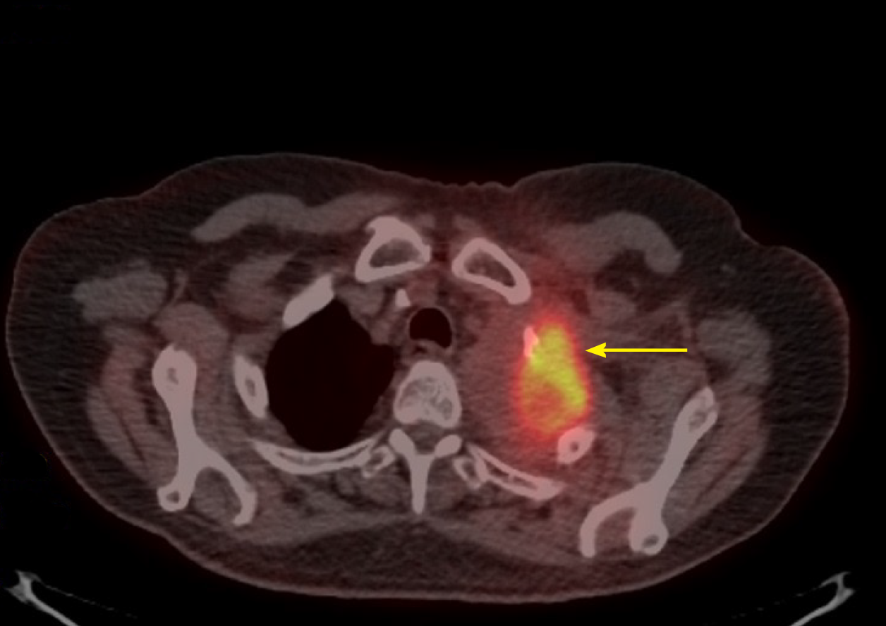
Figure 3 Chest positron emission tomography - computed tomography (lung window) after 9 cycles of pembrolizumab, with the yellow arrow pointing to an fluorodeoxyglucose-avid 5.
5 cm × 4 cm mass in the left upper lobe, with central necrosis and a maximum standardized uptake value of 8.6, consistent with malignancy. Inferiorly and in its continuation, there is a second lesion measuring 2.3 cm × 1.8 cm with a maximum standardized uptake value of 7.2, interpreted as a remnant from the initial tumor.
- Citation: Cimpeanu E, Ahmed J, Zafar W, DeMarinis A, Bardarov SS, Salman S, Bloomfield D. Pembrolizumab - emerging treatment of pulmonary sarcomatoid carcinoma: A case report. World J Clin Cases 2020; 8(1): 97-102
- URL: https://www.wjgnet.com/2307-8960/full/v8/i1/97.htm
- DOI: https://dx.doi.org/10.12998/wjcc.v8.i1.97


