Copyright
©The Author(s) 2020.
World J Clin Cases. Jan 6, 2020; 8(1): 200-207
Published online Jan 6, 2020. doi: 10.12998/wjcc.v8.i1.200
Published online Jan 6, 2020. doi: 10.12998/wjcc.v8.i1.200
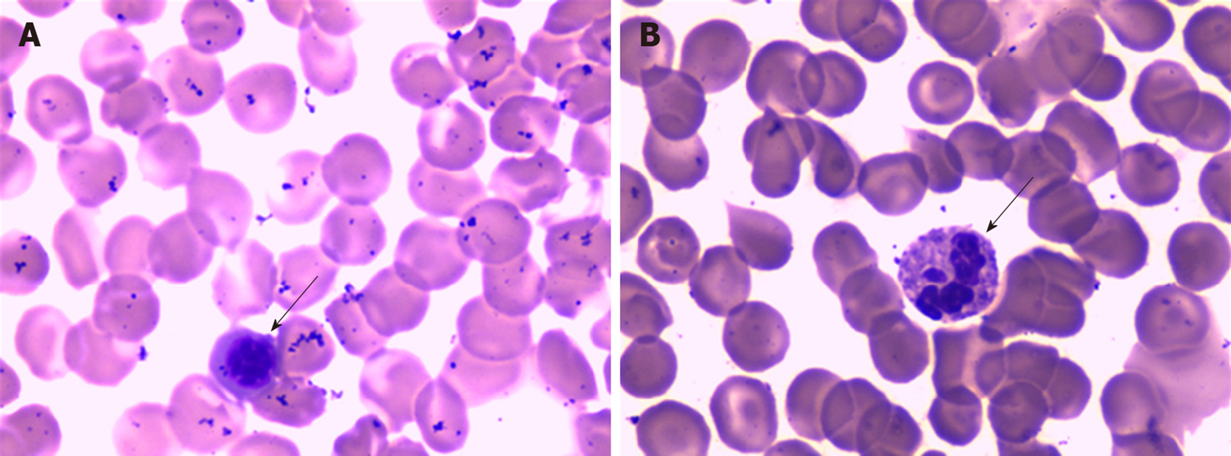
Figure 1 The peripheral blood smear showed a left shift of neutrophil nuclei, prominent toxic granulations and vacuolation in the neutrophil cytoplasm (see arrows) (Wright stain ×1000).
Figure 2 Computed tomography scan.
A: Head computed tomography (CT) scan; B: Multislice CT chest scan, showing little interstitial change in the lower lung; C: An abdominal CT scan suggested cardiac dysfunction with pulmonary edema.
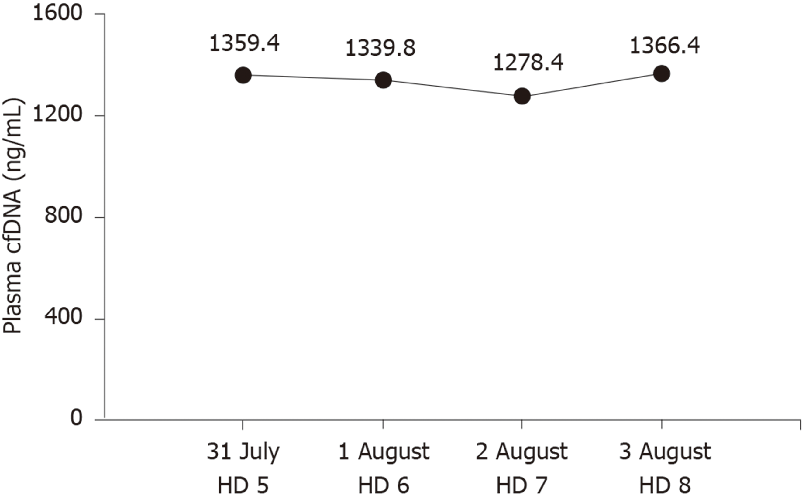
Figure 3 Plasma cell-free DNA concentrations.
cfDNA: Cell-free DNA; HD: Day of hospitalization.
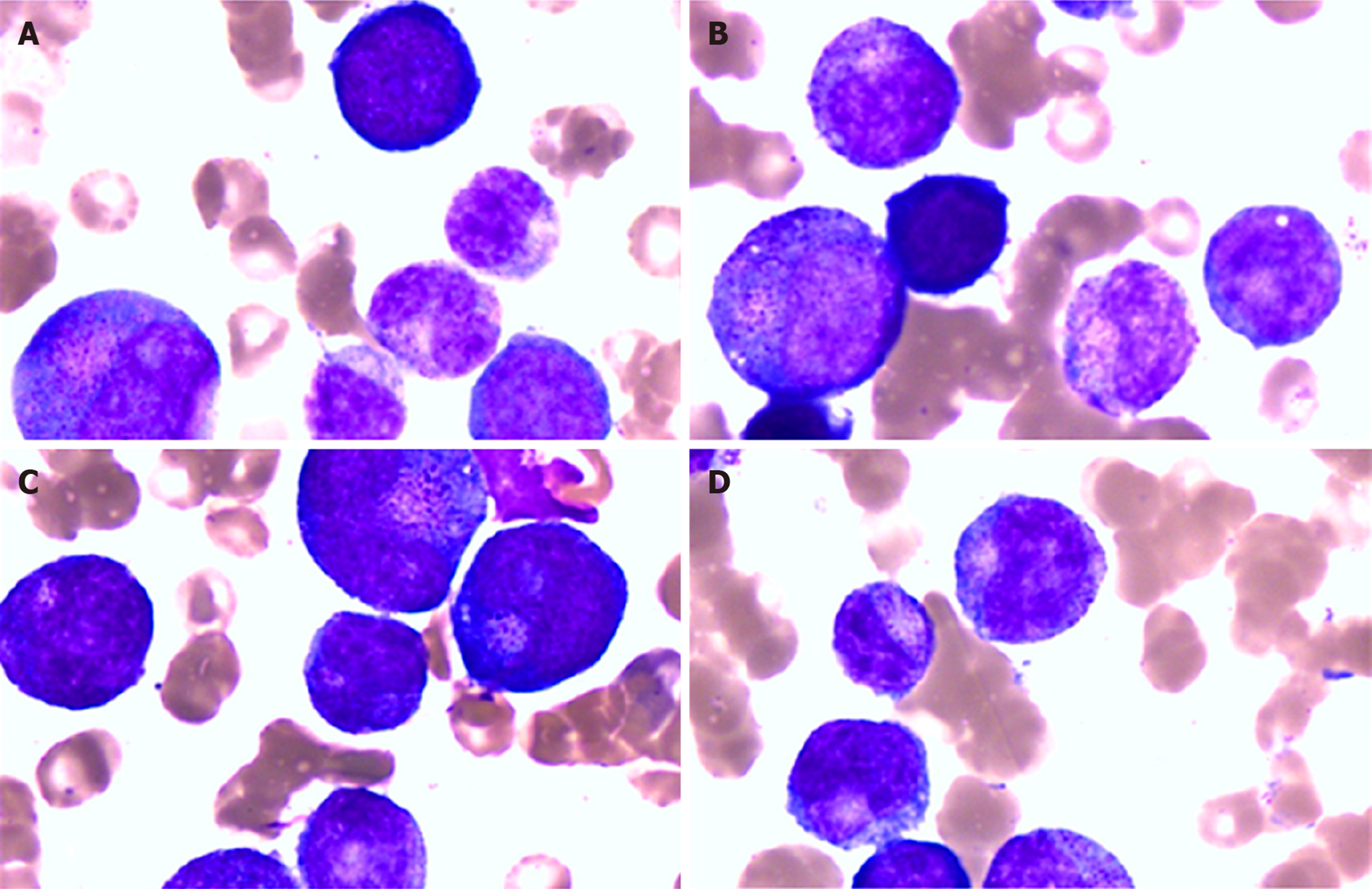
Figure 4 The bone marrow smear showed granulocyte maturation disorders.
A-D: Bone marrow smear. Wright stain ×1000.
- Citation: Liu JP, Zhang SC, Pan SY. Value of dynamic plasma cell-free DNA monitoring in septic shock syndrome: A case report. World J Clin Cases 2020; 8(1): 200-207
- URL: https://www.wjgnet.com/2307-8960/full/v8/i1/200.htm
- DOI: https://dx.doi.org/10.12998/wjcc.v8.i1.200


