Copyright
©The Author(s) 2020.
World J Clin Cases. Jan 6, 2020; 8(1): 168-174
Published online Jan 6, 2020. doi: 10.12998/wjcc.v8.i1.168
Published online Jan 6, 2020. doi: 10.12998/wjcc.v8.i1.168
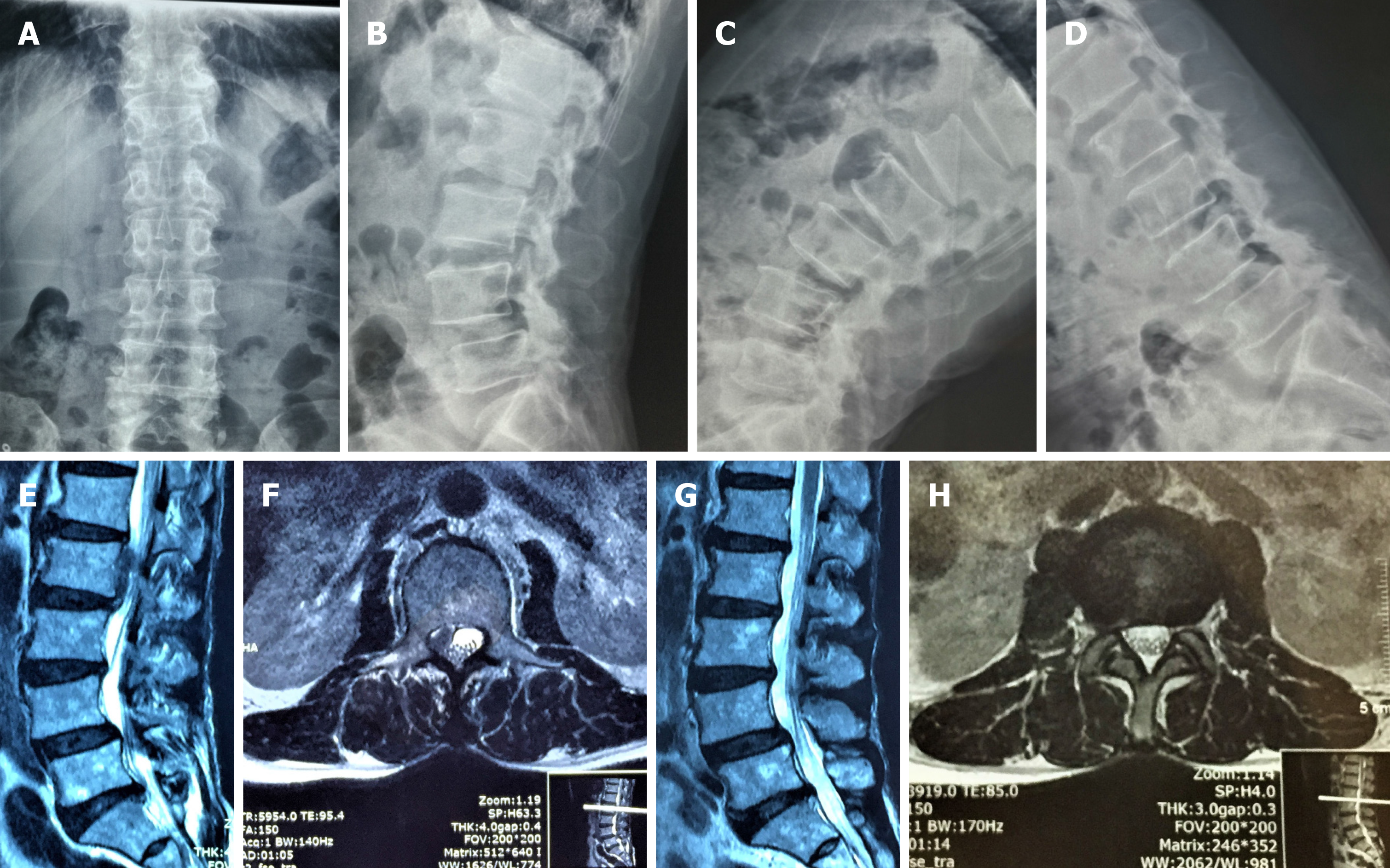
Figure 1 Preoperative and postoperative imaging examination.
A, B, C, D: Preoperative dynamic imaging x-ray examination indicated instability of the L4/5 vertebral body; E, F: Preoperative magnetic resonance imaging revealed L2/3 disc herniation with the nucleus pulposus highly migrating upward to the upper margin of the L2 vertebral body; G, H: Postoperative magnetic resonance imaging examination revealed clean removal of the nucleus pulposus.
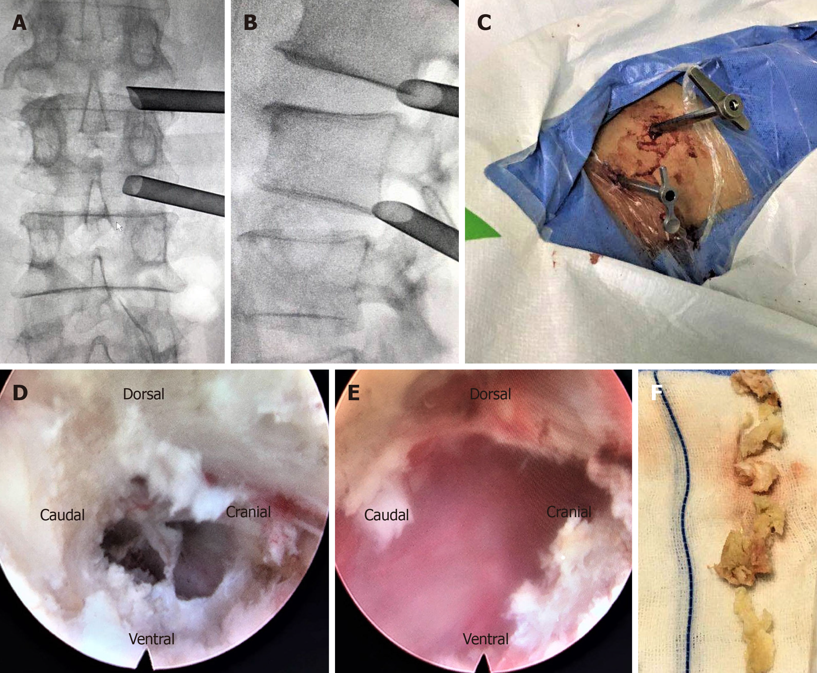
Figure 2 Surgical procedure of two-level percutaneous endoscopic lumbar discectomy.
A, B, C: Working channels placed into the intervertebral foramen at L1/2 and L2/3 level; D, E: Intraoperative endoscopic images of L1/2 and L2/3: complete removal of the nucleus pulposus; F: Removal of nucleus pulposus tissues.
- Citation: Wu XB, Li ZH, Yang YF, Gu X. Two-level percutaneous endoscopic lumbar discectomy for highly migrated upper lumbar disc herniation: A case report. World J Clin Cases 2020; 8(1): 168-174
- URL: https://www.wjgnet.com/2307-8960/full/v8/i1/168.htm
- DOI: https://dx.doi.org/10.12998/wjcc.v8.i1.168


