Copyright
©The Author(s) 2020.
World J Clin Cases. Jan 6, 2020; 8(1): 157-167
Published online Jan 6, 2020. doi: 10.12998/wjcc.v8.i1.157
Published online Jan 6, 2020. doi: 10.12998/wjcc.v8.i1.157
Figure 1 Enhanced total abdominal computed tomography.
A, B: Computed tomography scan showing that part of the descending colon, sigmoid colon (A), and rectum (B) were thickened with edema and numerous tiny dense spots were considered as phleboliths which were distributed within the intestinal wall.
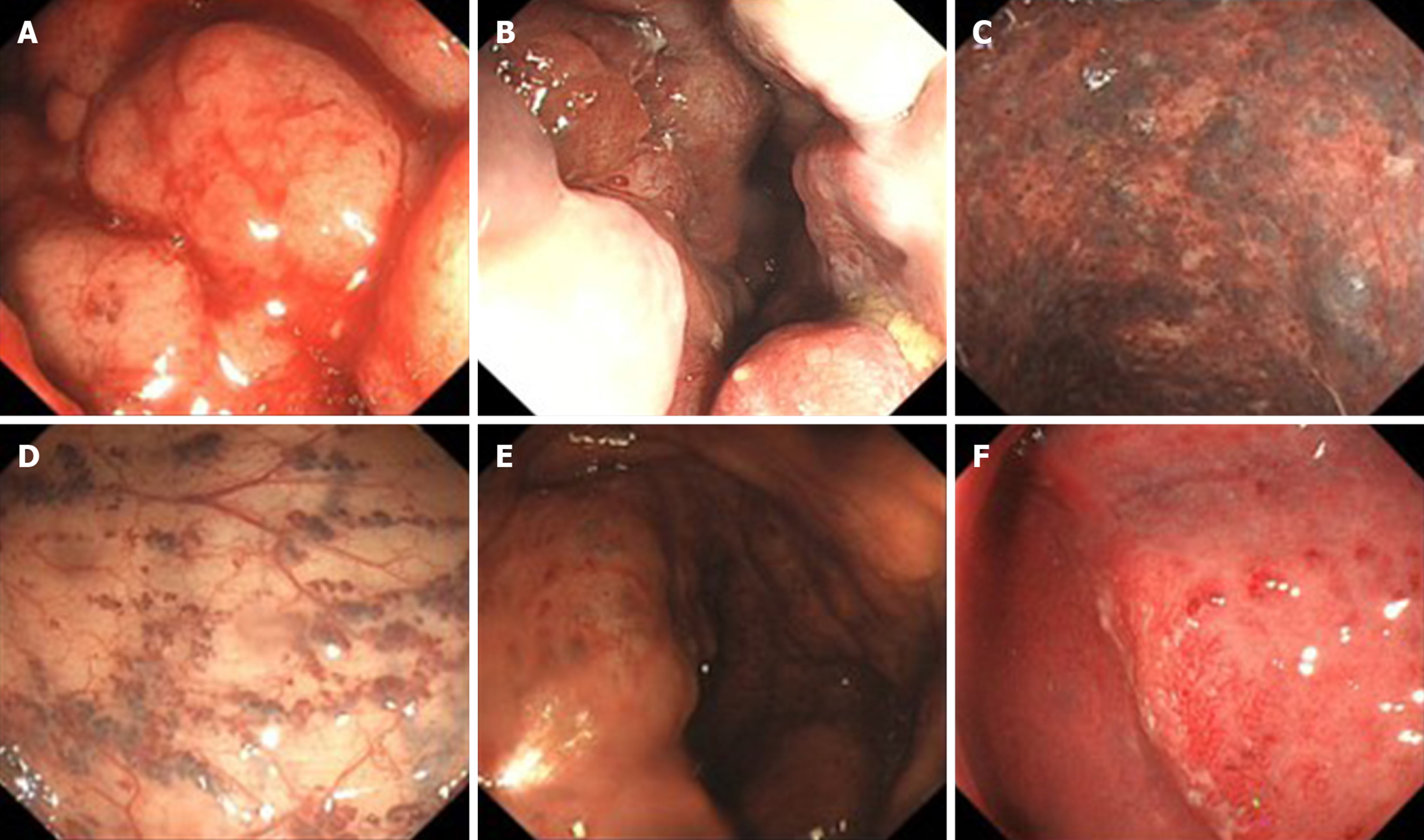
Figure 2 Colonoscopic findings.
A, B: Endoscopy displayed a pseudotumoral, smooth, embossed, vessel-like mass, which occupied the sigmoid colon and rectum; C: Bluish submucosal serpentine varicosities; D: Sporadic lamellar submucosal lesions; E: Ischemic appearance; F: Congestion, swelling and bleeding.
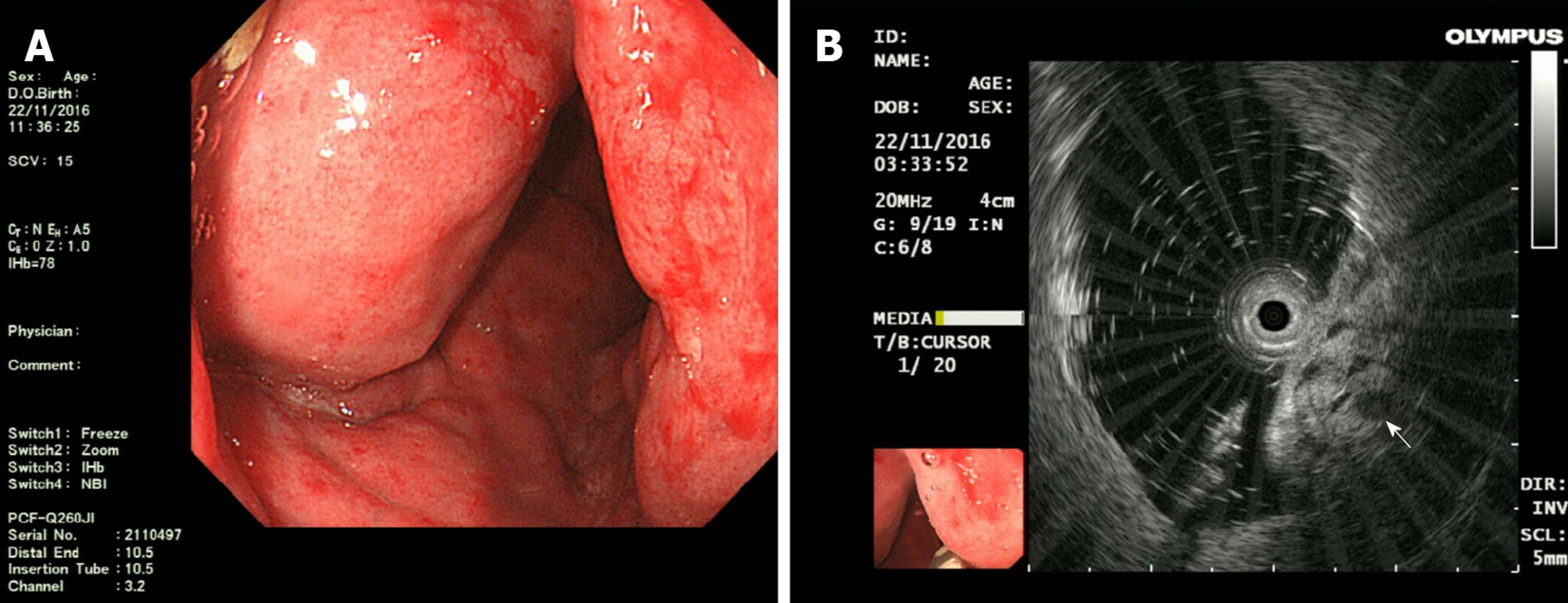
Figure 3 Endoscopic ultrasonography findings: A: Endoscopic ultrasonography (EUS) showed distinct edema and dense vascular varicose dilatations in the submucosal area with erosion and redness in the intestinal mucosa; B: The circumferential wall revealed multiple small “anechoic cystic spaces” on EUS (arrow).
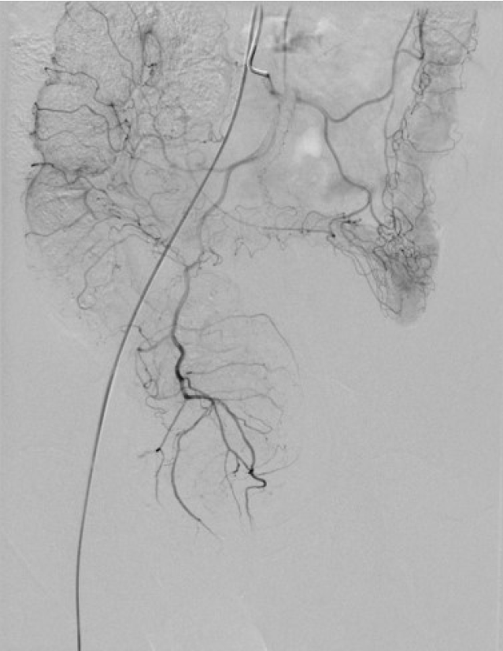
Figure 4 Superior rectal artery was normal and only part of the descending colon and rectosigmoid colon was thickened on angiography.
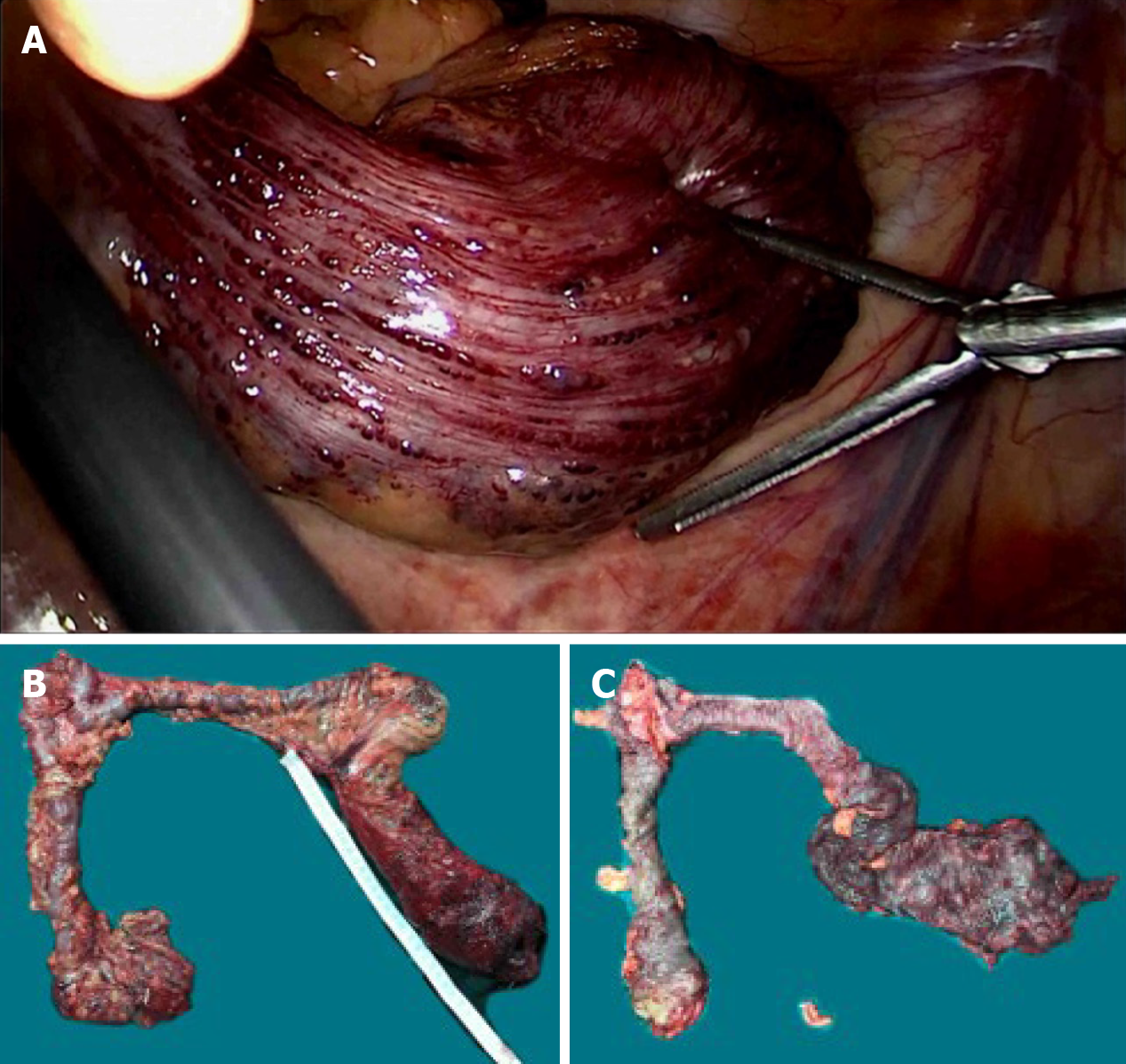
Figure 5 Intra-operative findings.
A: Vascular exophytic excrescences spread diffusely along the serosal surface of part of the thickened descending colon, sigmoid colon and rectum; B, C: Colon lesions were dark red in color with submucosal vascular tortuosity.
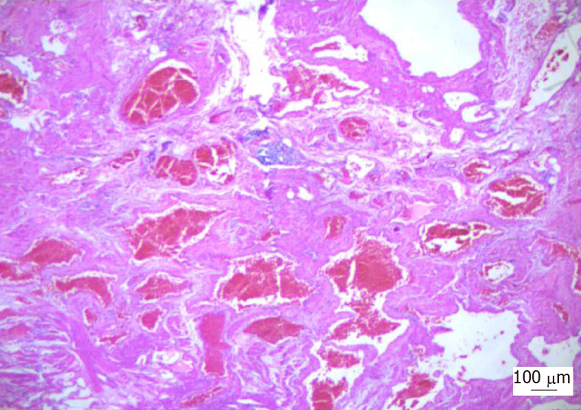
Figure 6 Photomicrograph of the resected specimen showed diffuse venous malformation lesions extending from the submucosa to the subserosa throughout the entire colorectum (× 100).
- Citation: Li HB, Lv JF, Lu N, Lv ZS. Mechanical intestinal obstruction due to isolated diffuse venous malformations in the gastrointestinal tract: A case report and review of literature. World J Clin Cases 2020; 8(1): 157-167
- URL: https://www.wjgnet.com/2307-8960/full/v8/i1/157.htm
- DOI: https://dx.doi.org/10.12998/wjcc.v8.i1.157


