Copyright
©The Author(s) 2020.
World J Clin Cases. Jan 6, 2020; 8(1): 140-148
Published online Jan 6, 2020. doi: 10.12998/wjcc.v8.i1.140
Published online Jan 6, 2020. doi: 10.12998/wjcc.v8.i1.140
Figure 1 Enteroscopy and segmental resection of primary and satellite jejunal lesions.
A: Primary discoid elevated lesion of 3 cm × 2 cm in the middle jejunum; B: Small satellite white nodule lesion 25 cm distal from the primary lesion; C: Photograph of the segmental jejunal resection including the primary and satellite lesions of lymphangioma.
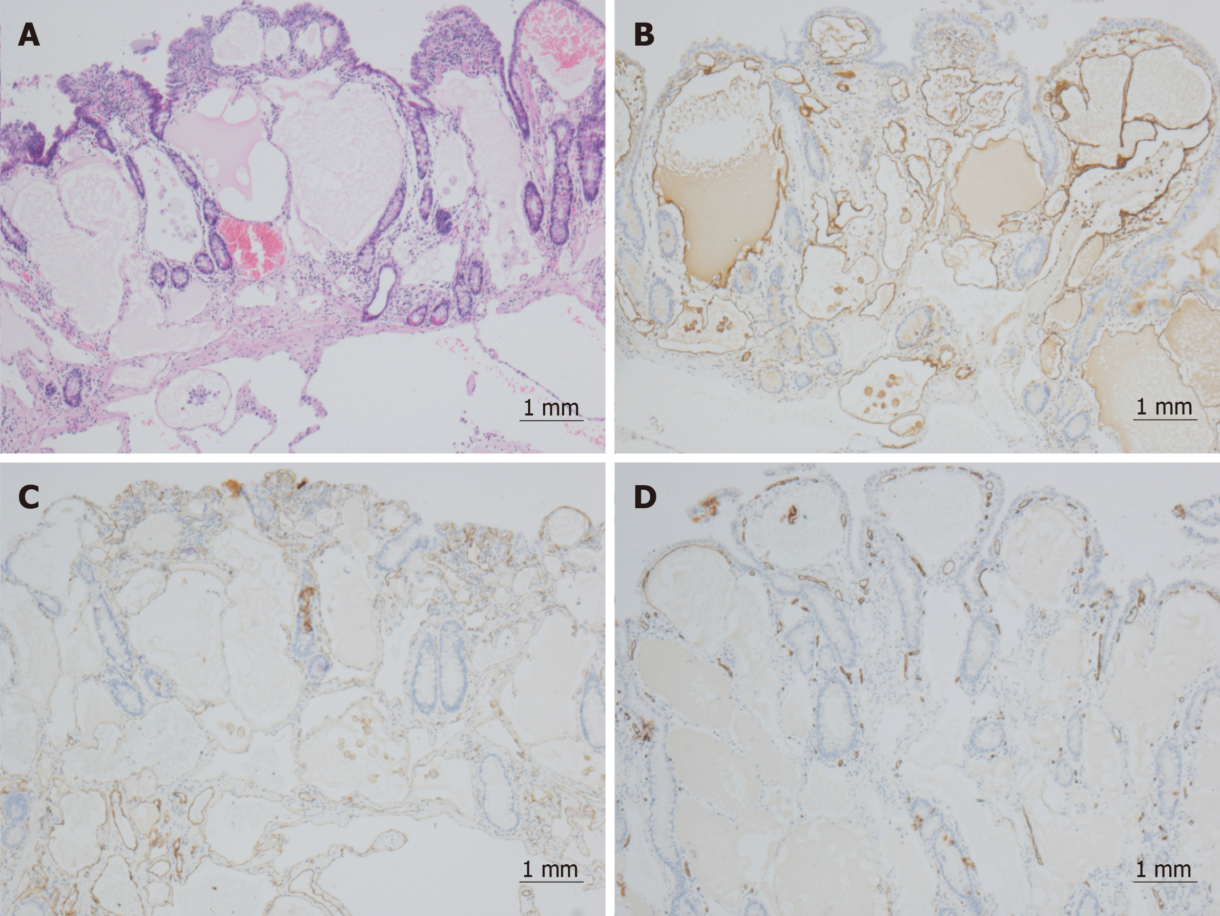
Figure 2 Microscopic features of the resected tumor.
A: Morphology showed variably sized cysts in mucosal lamina propria and submucosa, the lumen was filled with lymphatic fluid, and contained RBCs and lymphocytes (hematoxylin–eosin staining, 60×); B–D: Dilated cysts lined by a singer layer of flattened cells and immunohistochemical staining were positive for D2-40 (B) and CD31 (C), and negative for CD34 (D) (60×).
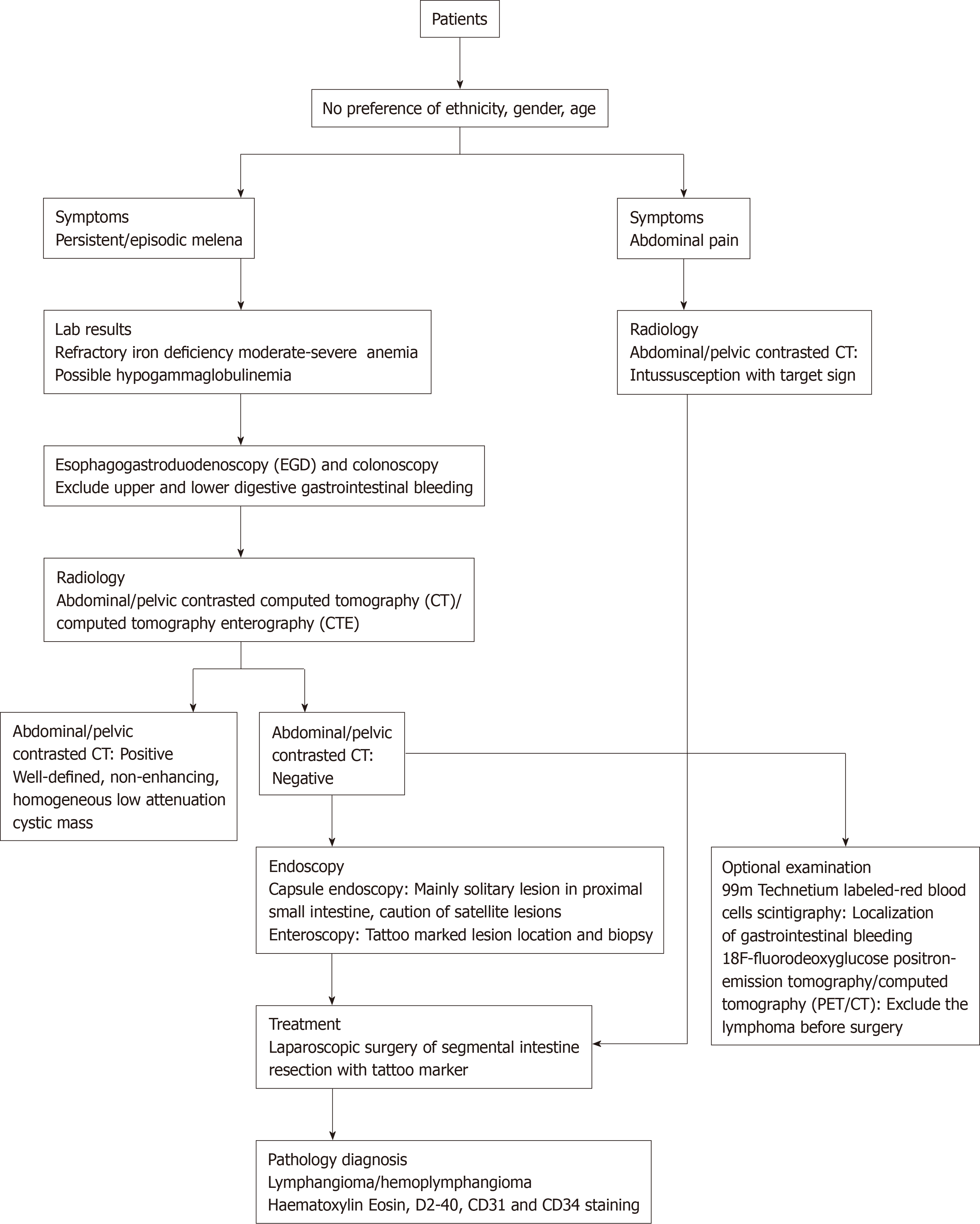
Figure 3 Recommended algorithm for identification and management of small intestine lymphangioma.
- Citation: Tan B, Zhang SY, Wang YN, Li Y, Shi XH, Qian JM. Jejunal cavernous lymphangioma manifested as gastrointestinal bleeding with hypogammaglobulinemia in adult: A case report and literature review. World J Clin Cases 2020; 8(1): 140-148
- URL: https://www.wjgnet.com/2307-8960/full/v8/i1/140.htm
- DOI: https://dx.doi.org/10.12998/wjcc.v8.i1.140


