Copyright
©The Author(s) 2019.
World J Clin Cases. Apr 6, 2019; 7(7): 881-890
Published online Apr 6, 2019. doi: 10.12998/wjcc.v7.i7.881
Published online Apr 6, 2019. doi: 10.12998/wjcc.v7.i7.881
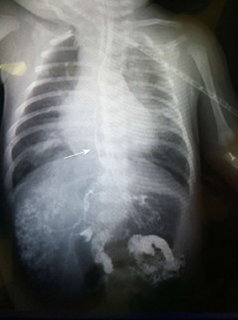
Figure 1 Computed tomography images.
A and B: Computed tomography (CT) scan and tracheoesophageal reconstruction; the arrow indicates the abnormal connection between the right bronchus and biliary tract. C: CT scan; the white arrow indicates gas in the biliary tract.
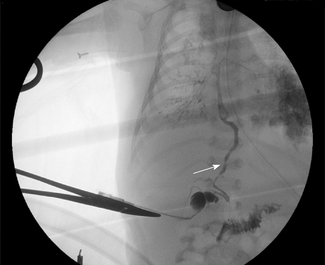
Figure 2 Contrast study via a fiberoptic bronchoscope.
The contrast dye can be seen in the fistula, biliary tract, and duodenum, and the white arrow indicates the fistula.
Figure 3 Intraoperative cholangiogram showing the connection between the biliary tract and right bronchus and patent distal bile duct; the white arrow indicates the biliary tract.
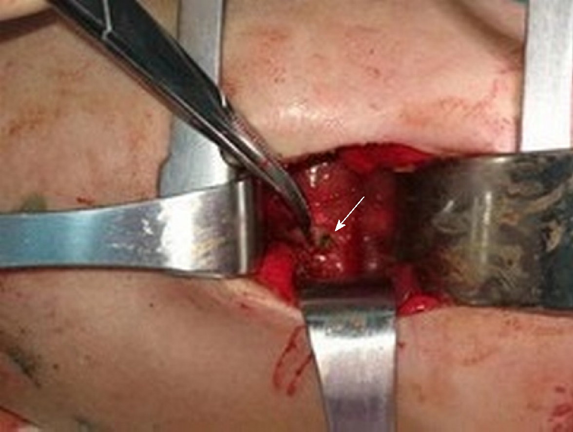
Figure 4 Methylene blue was injected into the gallbladder, and it flowed out from the puncture site; the white arrow indicates the puncture site.
Figure 5 A microscopic examination performed separately for three parts of the removed fistula.
A: The red arrow indicates the lumen that was partly covered by pseudo-ciliated columnar epithelium. The blue arrow indicates the cartilage rings that lay beneath the mucosa; B: The arrow indicates the clusters of bronchial glands at the end of the bronchus; C: The arrow indicates the glandular epithelia of the bile duct and interstitial cholestasis.
- Citation: Li TY, Zhang ZB. Congenital bronchobiliary fistula: A case report and review of the literature. World J Clin Cases 2019; 7(7): 881-890
- URL: https://www.wjgnet.com/2307-8960/full/v7/i7/881.htm
- DOI: https://dx.doi.org/10.12998/wjcc.v7.i7.881


