Copyright
©The Author(s) 2019.
World J Clin Cases. Apr 6, 2019; 7(7): 872-880
Published online Apr 6, 2019. doi: 10.12998/wjcc.v7.i7.872
Published online Apr 6, 2019. doi: 10.12998/wjcc.v7.i7.872
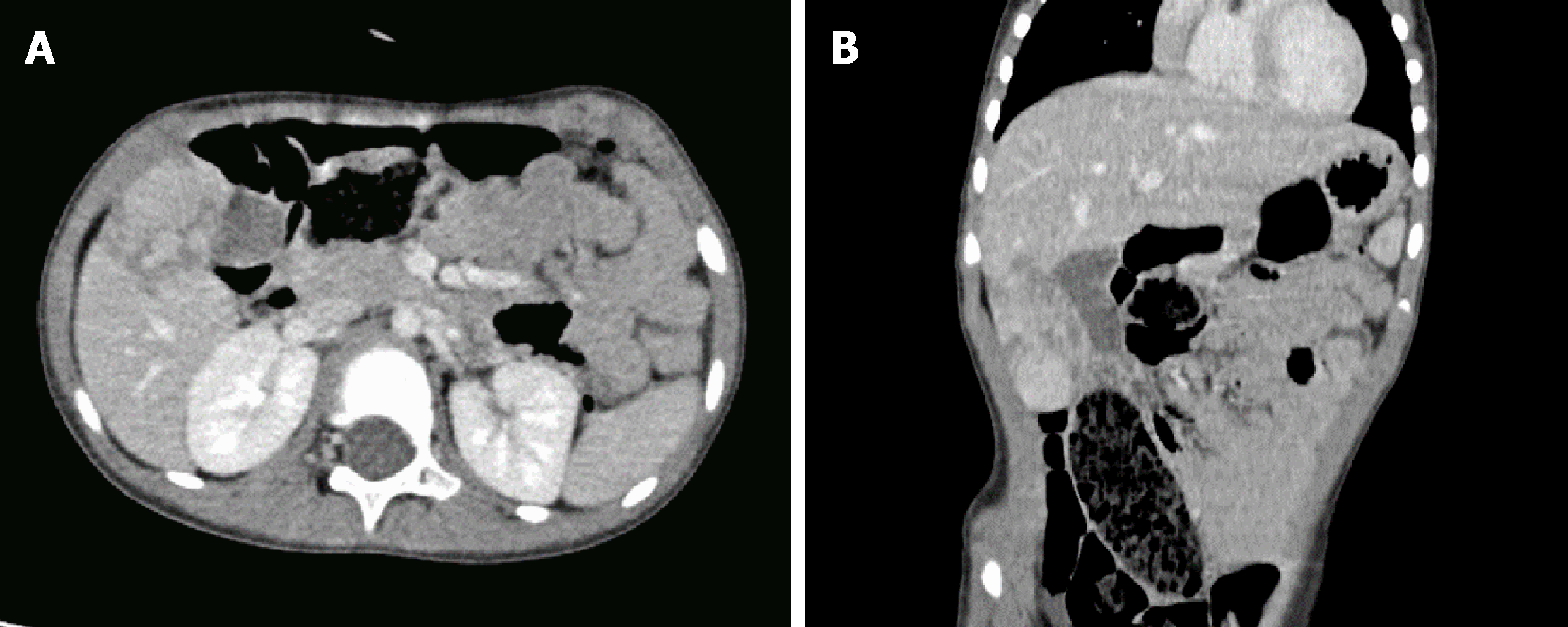
Figure 1 Contrast-enhanced computed tomography images revealing that the tumour was located in the 5th segment of the liver and was closely related to the gallbladder.
A: Axial plane; B: Coronal plane.
Figure 2 Preoperative 3-dimensional reconstruction images.
The relationship between the tumour and the hepatic vein is shown. A: Preoperational 3-dimensional reconstruction image showing the location of the tumour and gallbladder; B: Relationship between hepatic veins and the tumour; C: Image through the Cantlie line showing tumour’s drainage to the right and middle hepatic vein; D: Several branches of portal veins supplying blood to the tumour.
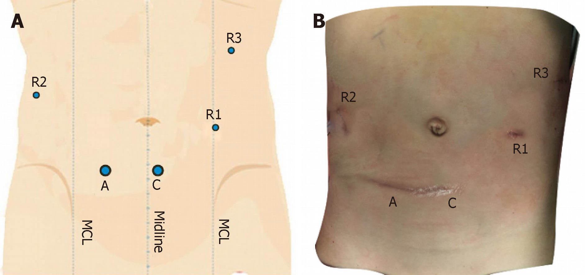
Figure 3 Schematic diagram (A) and actual picture (B) of trocar placement.
Postoperative cosmetic status is shown in B. A: Accessory port; C: Camera port: R1: No. 1 robotic arm; R2: No. 2 robotic arm; R3: No.3 robotic arm; MCL: Midclavicular line.
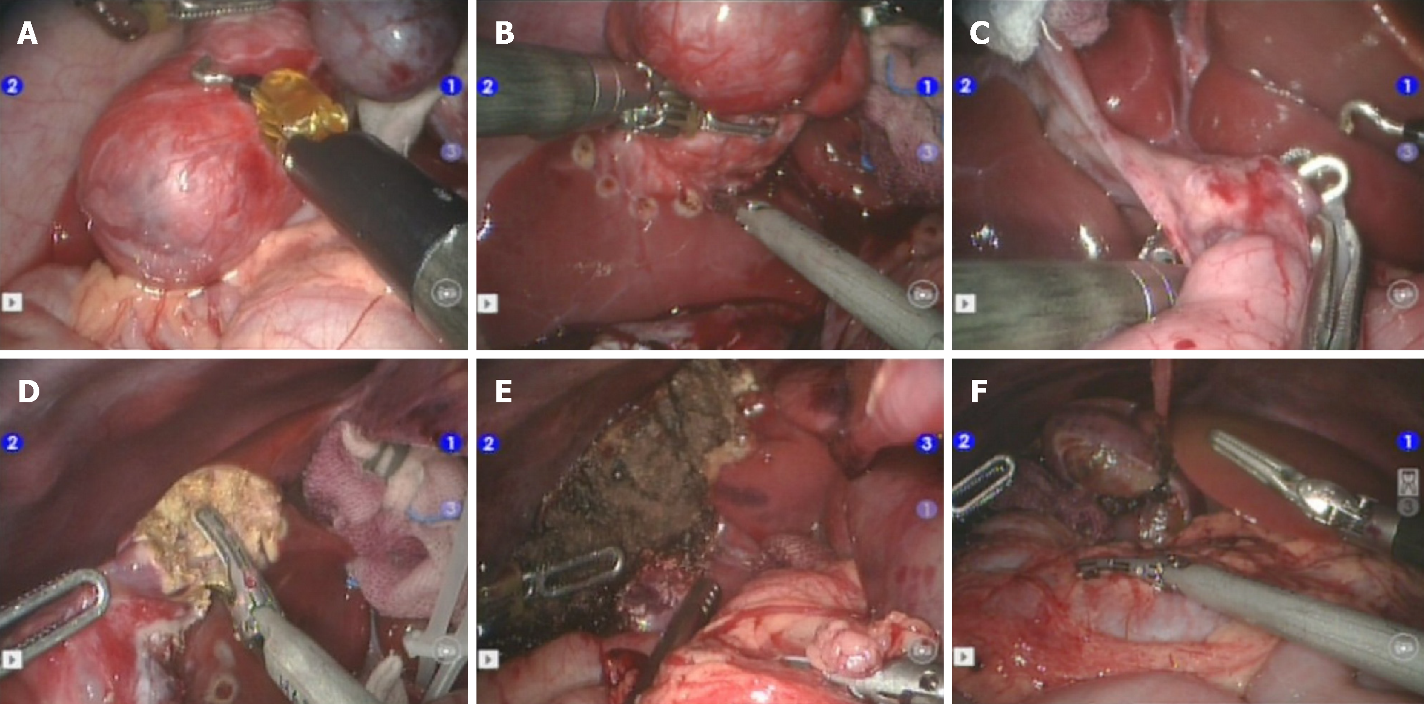
Figure 4 Surgery-related images.
A: Freeing of the gallbladder; B: Pre-cutting line location; C: Liver hilum blocking; D: Isolation of liver parenchyma; E: Liver cross section; F: Placement of the gallbladder naturally.
Figure 5 Excised specimen.
The tumour was radically resected and around 46 mm × 26 mm × 58 mm in size, without the gallbladder.
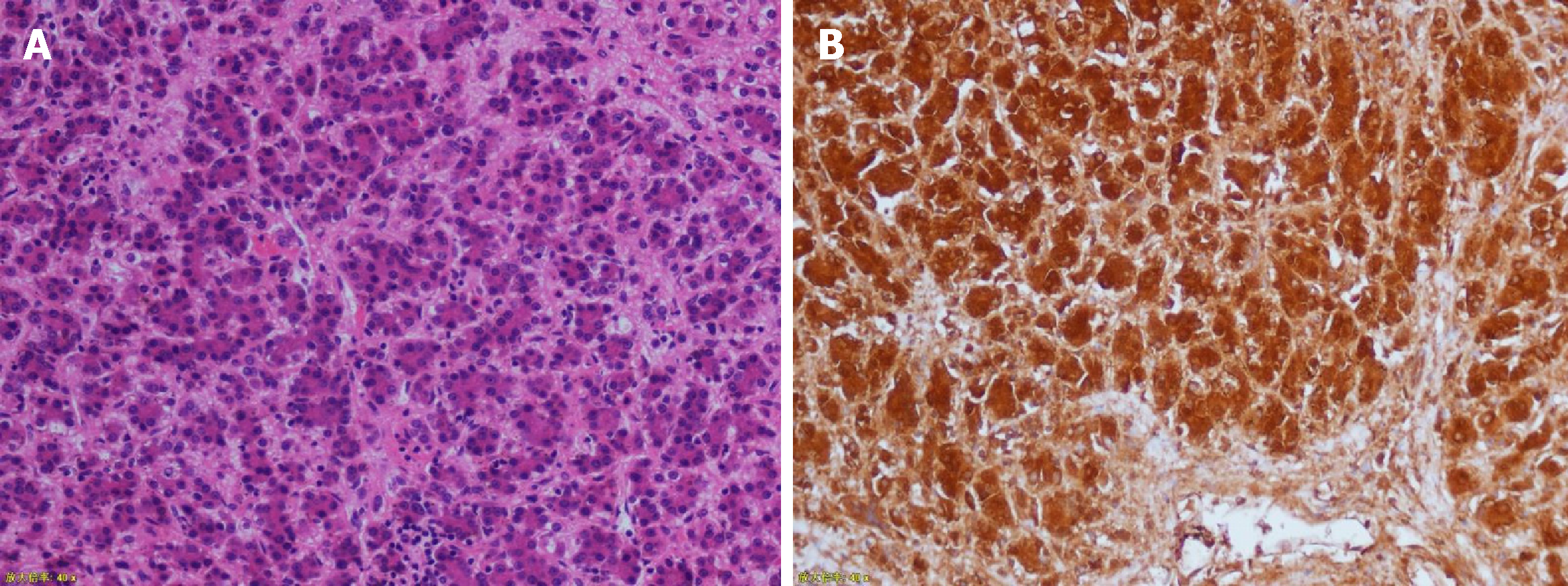
Figure 6 Pathological images (magnification 200×).
The immunohistochemical results were hepatocytes (+), GPC-3 (+), alpha fetoprotein (focal +), ki-67 (+20%), Arg-1 (+), and CD99 (local).
- Citation: Chen DX, Wang SJ, Jiang YN, Yu MC, Fan JZ, Wang XQ. Robot-assisted gallbladder-preserving hepatectomy for treating S5 hepatoblastoma in a child: A case report and review of the literature. World J Clin Cases 2019; 7(7): 872-880
- URL: https://www.wjgnet.com/2307-8960/full/v7/i7/872.htm
- DOI: https://dx.doi.org/10.12998/wjcc.v7.i7.872


