Copyright
©The Author(s) 2019.
World J Clin Cases. Apr 6, 2019; 7(7): 863-871
Published online Apr 6, 2019. doi: 10.12998/wjcc.v7.i7.863
Published online Apr 6, 2019. doi: 10.12998/wjcc.v7.i7.863
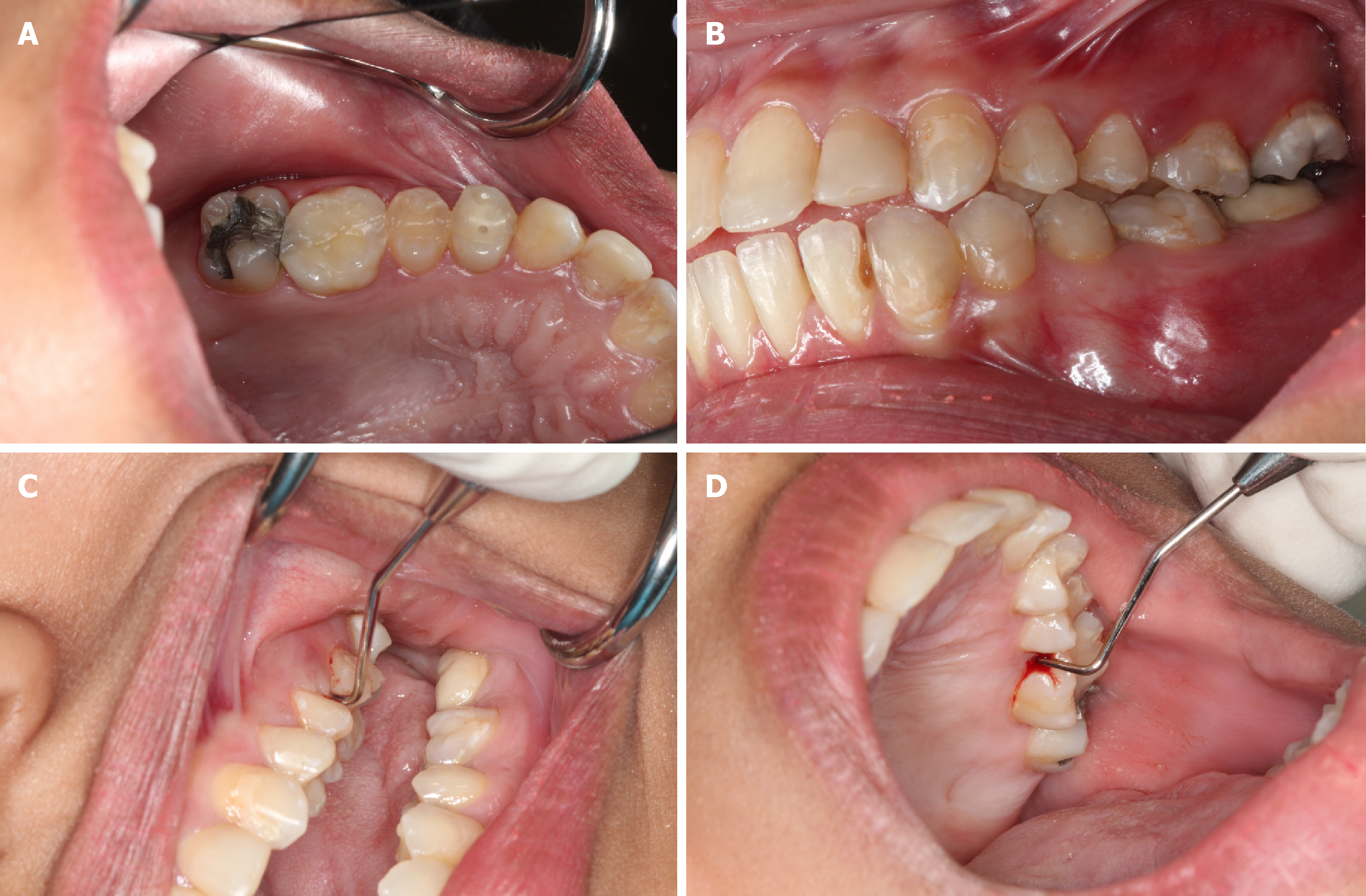
Figure 1 Clinical findings.
A, B: Localized erythematous swelling at the buccal interdental papilla between the left maxillary first molar and second premolars; C, D: Pocket depth of 12 mm and 10 mm in the mesiobuccal and mesiolingual regions of the left maxillary first molar.
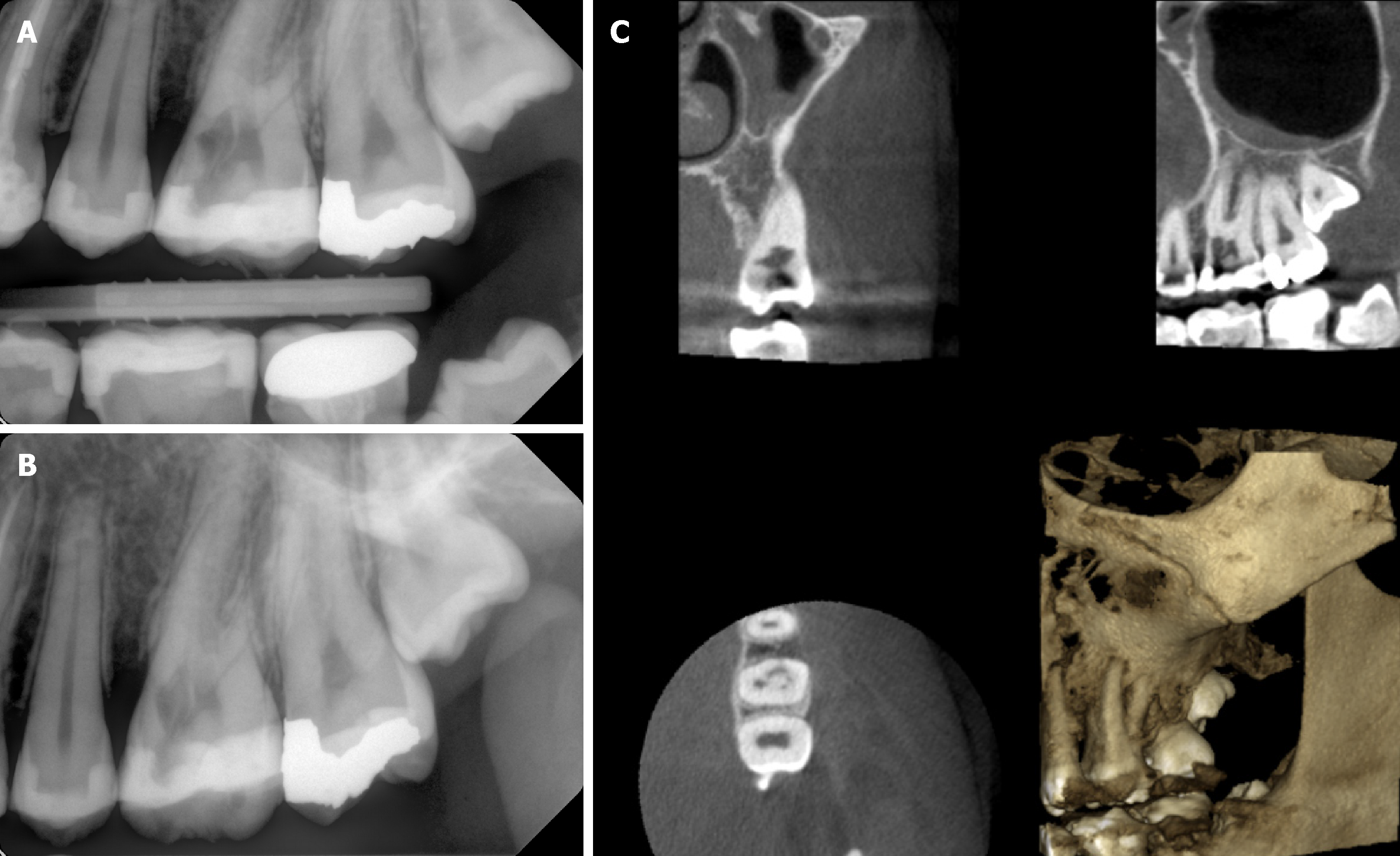
Figure 2 Radiographic findings.
A, B: Two dimensional radiographs revealed breakdown of alveolar crest mesial to the left maxillary first molar and irregular molted radiolucency mesial to the pulp space of the left maxillary first molar separated by radiopaque line and extending into the radicular dentin; C: Cone-beam computed tomographic images from different planes demonstrated the size and external perforation of the resorption defect.
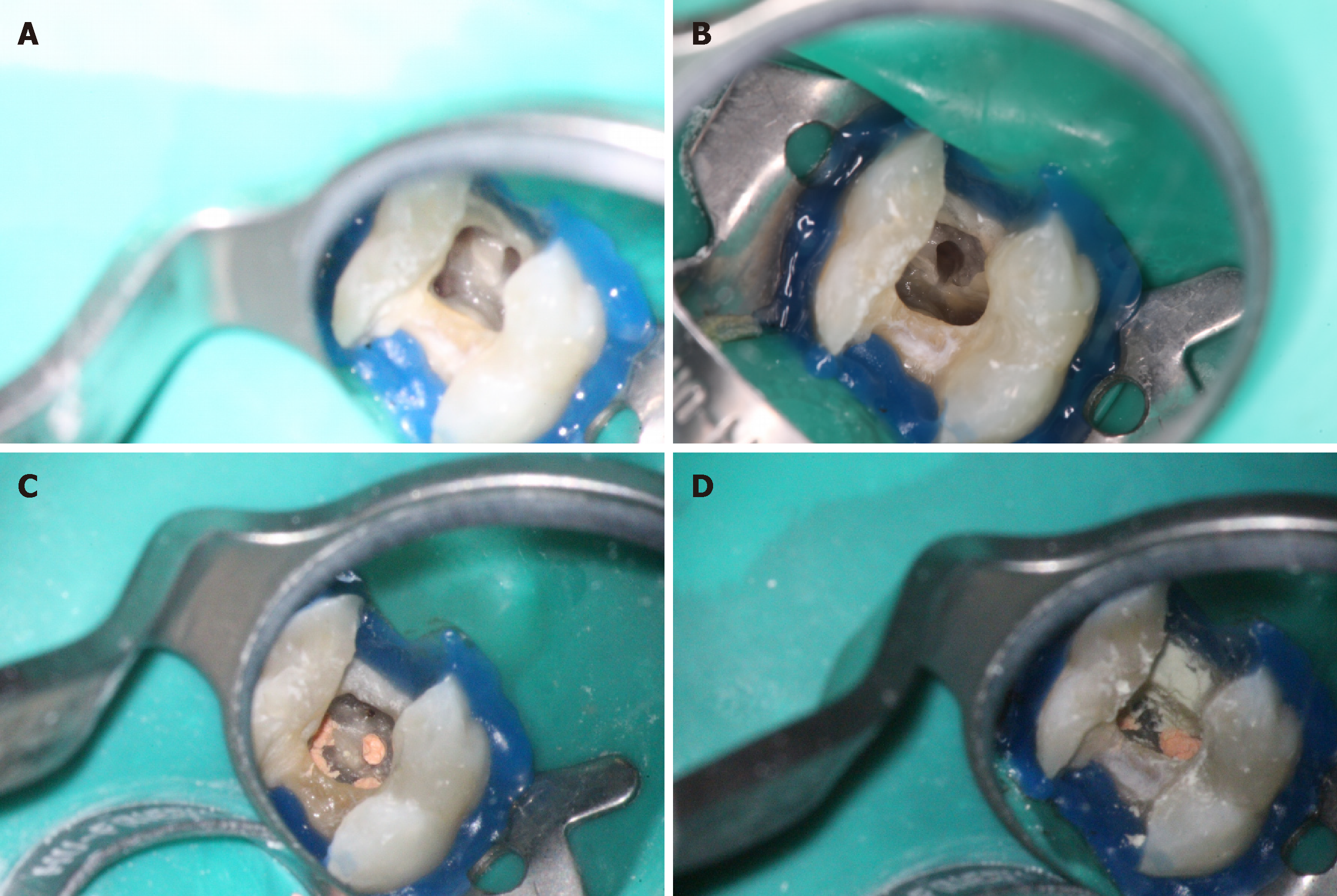
Figure 3 Second appointment.
A, B: Control of bleeding from the resorption defect and achievement of better visibility; C: Obturation of the root canal system using continuous wave of vertical condensation; D: resorption defect was sealed using Mineral trioxide aggregate.
Figure 4 Immediate postoperative radiograph: Adequate mineral trioxide aggregate filling of the perforation and resorption defect.
Figure 5 One-year recall: Probing depths around the tooth within normal limits of 3-4 mm and restoration of the epithelial attachment and crestal bone.
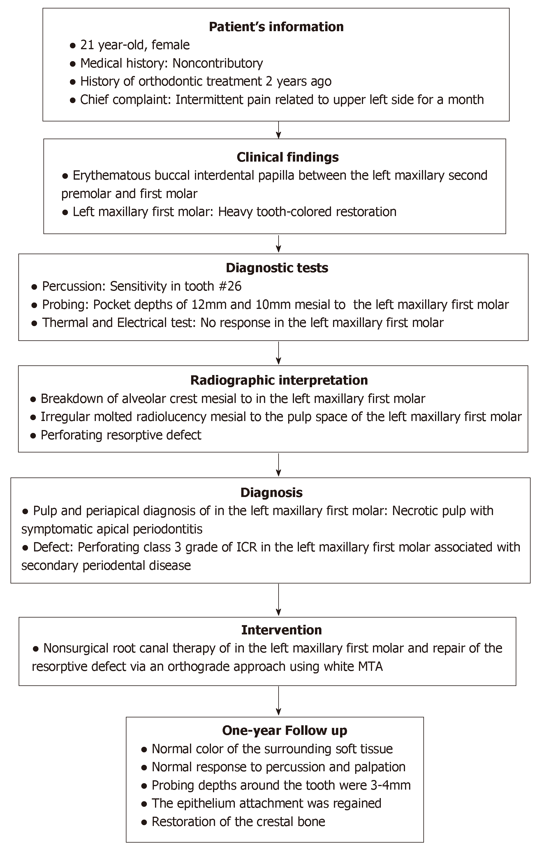
Figure 6 Timeline summarizing the patient’s information, clinical findings, diagnostic tests, diagnosis, intervention, and follow up.
- Citation: Alqedairi A. Non-Invasive management of invasive cervical resorption associated with periodontal pocket: A case report. World J Clin Cases 2019; 7(7): 863-871
- URL: https://www.wjgnet.com/2307-8960/full/v7/i7/863.htm
- DOI: https://dx.doi.org/10.12998/wjcc.v7.i7.863


