Copyright
©The Author(s) 2019.
World J Clin Cases. Mar 6, 2019; 7(5): 656-662
Published online Mar 6, 2019. doi: 10.12998/wjcc.v7.i5.656
Published online Mar 6, 2019. doi: 10.12998/wjcc.v7.i5.656
Figure 1 Preoperative images of the tumor.
A: The paraganglioma located in the carotid artery bifurcation (red arrow); B: A low-density nodule of the thyroid isthmus with a high-density calcification can be seen (blue arrow); C: Enhanced computed tomography showing coexistence of paraganglioma (red arrow) and thyroid carcinoma (blue arrow).
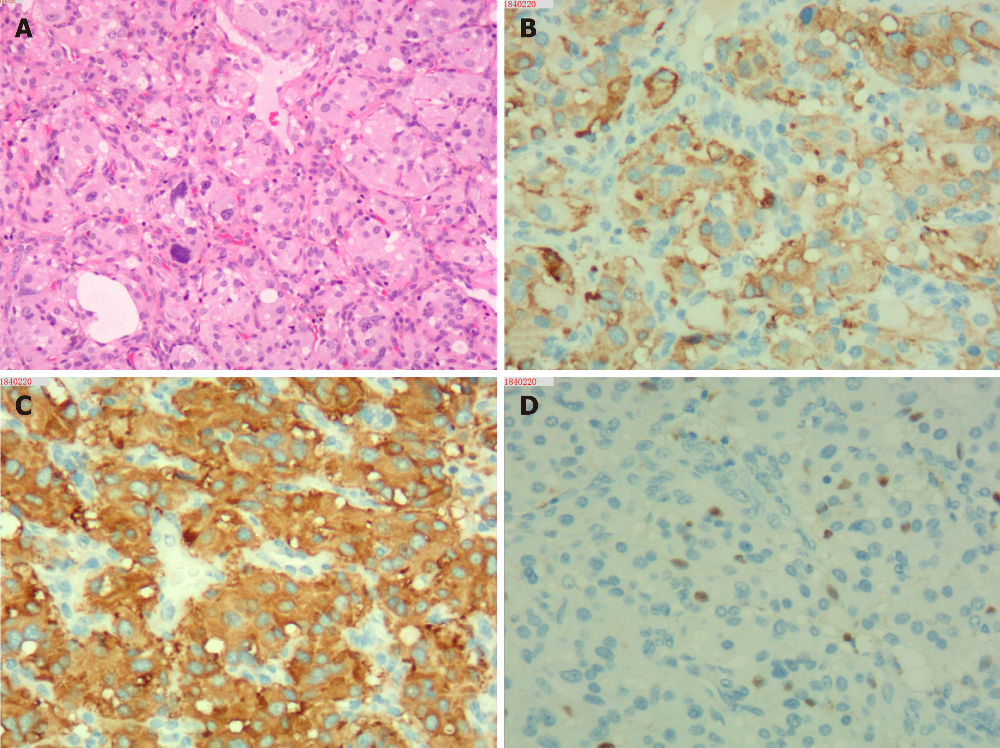
Figure 2 Microscopic features of the paraganglioma.
A: Hematoxylin-Eosin (HE) staining (100 ×) showing well-defined solid nests of tumor cells, rounded by a fibrovascular tissue; B-C: Immunohistochemical staining of the tumor showing that the chief cells are intensively positive for chromogranin (B) and synaptophysin (C); D: Sustentacular cells are positive for S-100 protein.
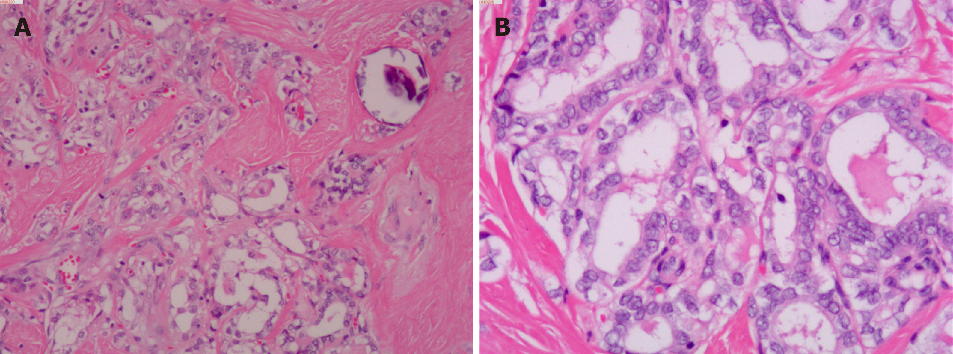
Figure 3 Microscopic features of the papillary thyroid carcinoma.
A: Hematoxylin-Eosin (HE) staining (40 ×) showing that the papillae in papillary thyroid carcinoma are composed of cuboidal cells; B: HE staining (100 ×) showing nuclear changes including nuclear clearing and overlapping nuclei.
- Citation: Lin B, Yang HY, Yang HJ, Shen SY. Concomitant paraganglioma and thyroid carcinoma: A case report. World J Clin Cases 2019; 7(5): 656-662
- URL: https://www.wjgnet.com/2307-8960/full/v7/i5/656.htm
- DOI: https://dx.doi.org/10.12998/wjcc.v7.i5.656


