Copyright
©The Author(s) 2019.
World J Clin Cases. Feb 26, 2019; 7(4): 532-537
Published online Feb 26, 2019. doi: 10.12998/wjcc.v7.i4.532
Published online Feb 26, 2019. doi: 10.12998/wjcc.v7.i4.532
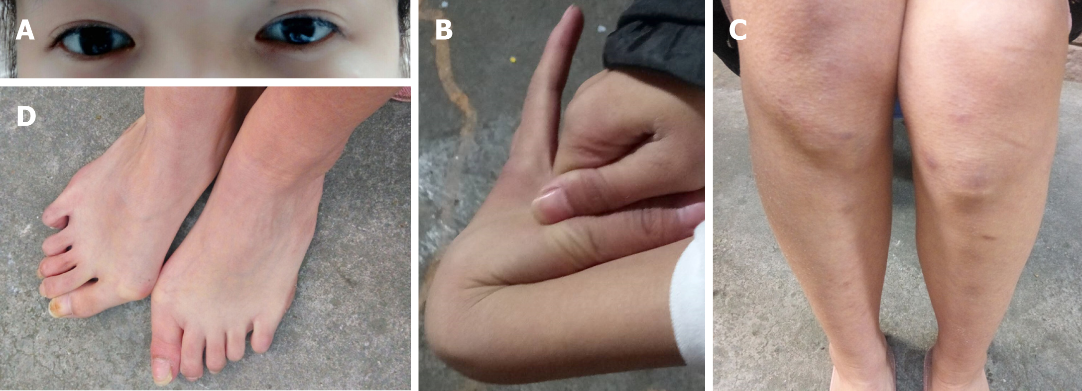
Figure 1 Clinical features of the girl with Ehlers-Danlos syndrome VI.
A: Blue tint to the sclera; B: Joint hypermobility; C: Abnormal scar; D: Planovalgus feet.
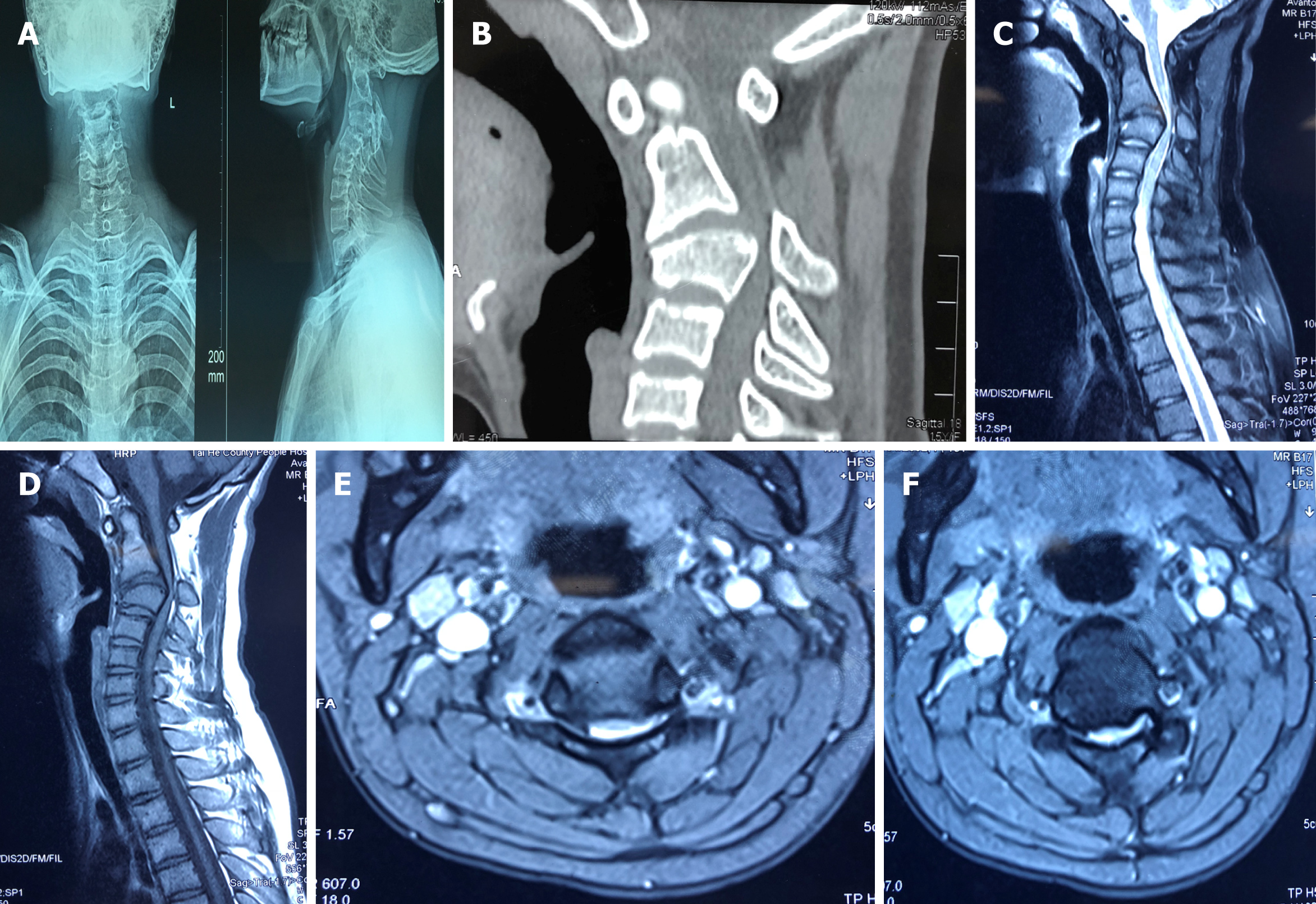
Figure 2 Preoperative imaging of a 16-year-old-female with Ehlers-Danlos syndrome VI.
X-ray (A) and computed tomography (B) images demonstrating high cervical kyphosis from C2 to C4. Magnetic resonance imaging (C-F) showing that the cervical ventral spinal cord was compressed by the C3 vetebra.
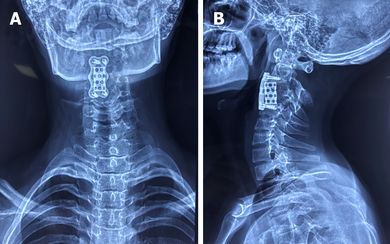
Figure 3 Follow-up X-ray images.
A: Antero-posterior view radiograph of the cervical spine; B: Lateral view radiograph of the cervical spine. Radiographs acquired 6 wk following anterior cervical corpectomy decompression and fusion showing that the preoperative kyphotic deformity in the cervical spine was significantly corrected.
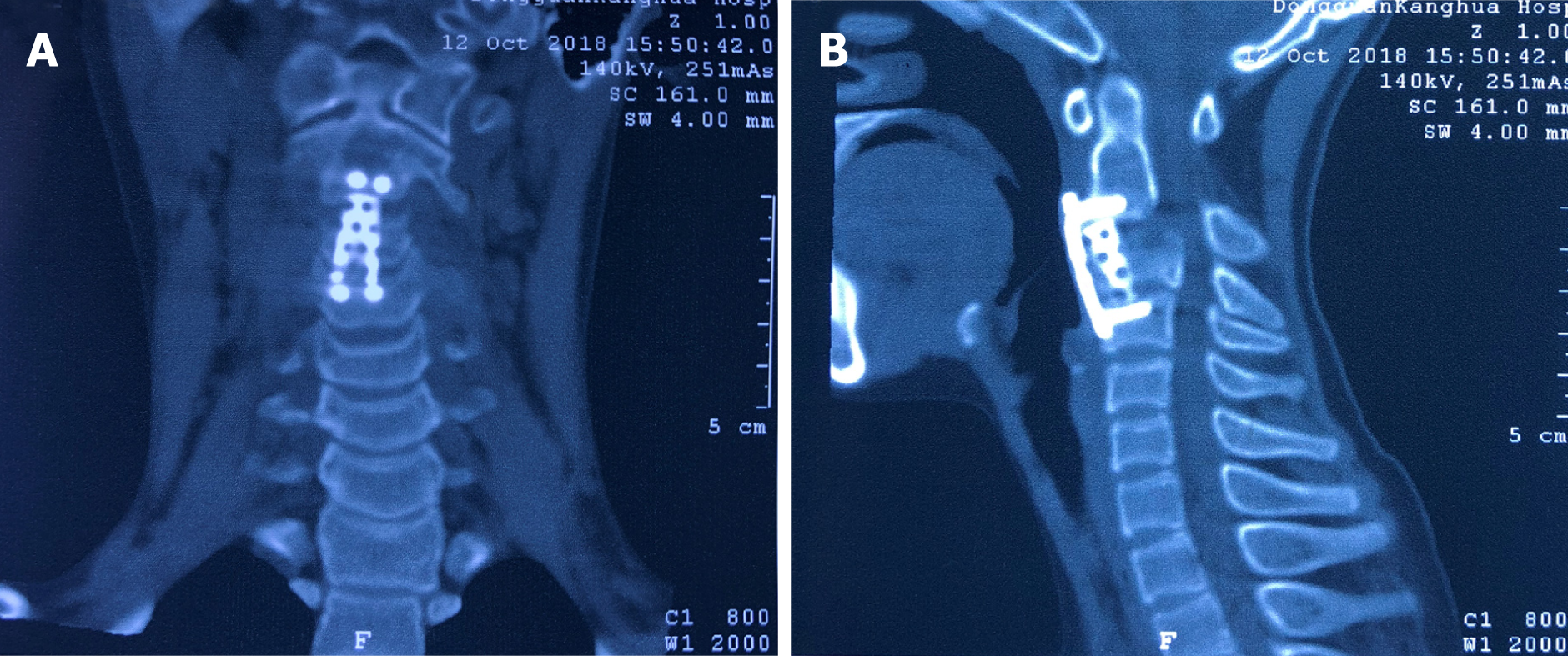
Figure 4 Follow-up computed tomography images.
Computed tomography acquired 5 mo following surgery with images in the coronal (A) plane and sagittal reconstruction (B) showing maintenance of alignment and evidence of a solid arthrodesis.
- Citation: Fang H, Liu PF, Ge C, Zhang WZ, Shang XF, Shen CL, He R. Anterior cervical corpectomy decompression and fusion for cervical kyphosis in a girl with Ehlers-Danlos syndrome: A case report. World J Clin Cases 2019; 7(4): 532-537
- URL: https://www.wjgnet.com/2307-8960/full/v7/i4/532.htm
- DOI: https://dx.doi.org/10.12998/wjcc.v7.i4.532


