Copyright
©The Author(s) 2019.
World J Clin Cases. Feb 26, 2019; 7(4): 419-430
Published online Feb 26, 2019. doi: 10.12998/wjcc.v7.i4.419
Published online Feb 26, 2019. doi: 10.12998/wjcc.v7.i4.419
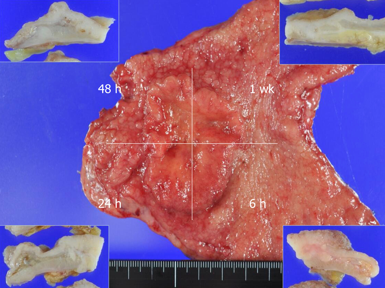
Figure 1 Representative image of a resected gastric cancer specimen.
Each patient’s resected specimen was immediately divided into four pieces that were individually fixed in a solution for 6 h, 24 h, or 48 h or 1 wk. Insets: Cut surfaces of the specimen after fixation for each duration.
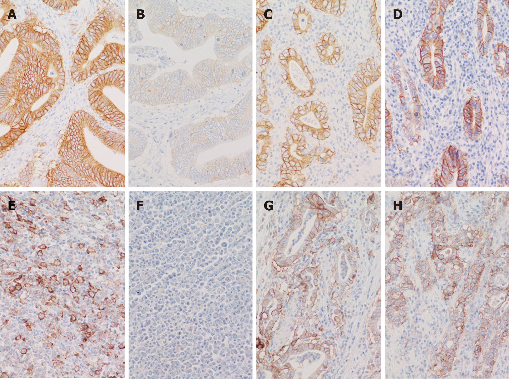
Figure 2 Representative immunohistochemistry images from very-long-fixation cases.
A, B: Human epidermal growth factor receptor 2 (HER-2) immunohistochemistry (IHC) of case 12. Diffuse and strong HER-2 expression (score 3+) was observed in the specimen piece fixed by 10% formalin for 1 week (A). The HER-2 expression was significantly weakened (assessed as 2+) after very-long fixation (19 mo) by 10% formalin (B); C, D: HER-2 IHC of case 31. Diffuse and strong HER-2 expression (score 3+) was observed in the piece fixed by 10% NBF for 1 wk (C), and this expression was maintained (score 3+) even after very-long fixation (16 mo) by 10% NBF (D); E, F: PD-L1 IHC of case 20. The PD-L1 expression by tumor cells was observed in the piece that underwent 24-h fixation by 10% formalin (E) but it completely disappeared after very-long fixation (28 mo) by 10% formalin (F); G, H: PD-L1 IHC of case 30. The PD-L1 expression by tumor cells was observed in the piece that underwent 1-wk fixation by 10% NBF (G) and was maintained even after very-long fixation (16 mo) by 10% NBF (H).
- Citation: Kai K, Yoda Y, Kawaguchi A, Minesaki A, Iwasaki H, Aishima S, Noshiro H. Formalin fixation on HER-2 and PD-L1 expression in gastric cancer: A pilot analysis using the same surgical specimens with different fixation times. World J Clin Cases 2019; 7(4): 419-430
- URL: https://www.wjgnet.com/2307-8960/full/v7/i4/419.htm
- DOI: https://dx.doi.org/10.12998/wjcc.v7.i4.419


