Copyright
©The Author(s) 2019.
World J Clin Cases. Feb 6, 2019; 7(3): 340-346
Published online Feb 6, 2019. doi: 10.12998/wjcc.v7.i3.340
Published online Feb 6, 2019. doi: 10.12998/wjcc.v7.i3.340
Figure 1 Pre-operative imaging examination.
A: Superior abdominal enhanced computed tomography (arterial phase); B: Abdominal magnetic resonance imaging (axial); C: Abdominal magnetic resonance imaging (coronal). T: Tumor; L: Liver; K: Kidney; P: Pancreas; IVC: Inferior vena cava.
Figure 2 Post-operative macroscopic examination of the tumor.
The tumor was 200 mm × 160 mm in diameter.
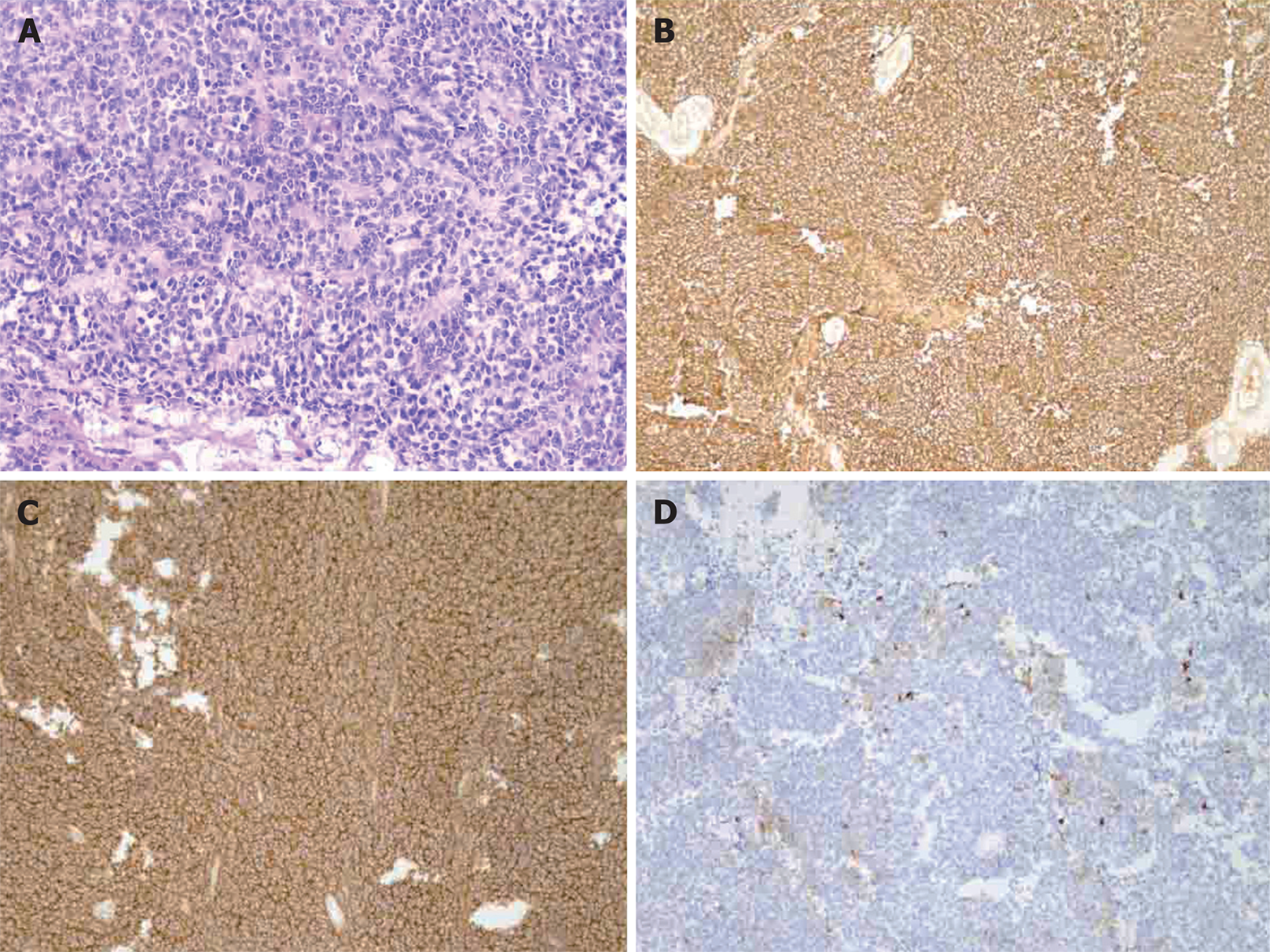
Figure 3 Pathological changes of the tumor.
A: Hematoxylin and eosin staining revealed small round tumor cells and several lobulated structures (Homer–Wright rosettes) (200 ×); B: Immunohistochemistry showed tumor cells were positive for CD99 (100 ×); C: Immunohistochemistry showed tumor cells were positive for neuron-specific enolase (100 ×); D: Immunohistochemistry showed tumor cells were positive for synaptophysin (100 ×).
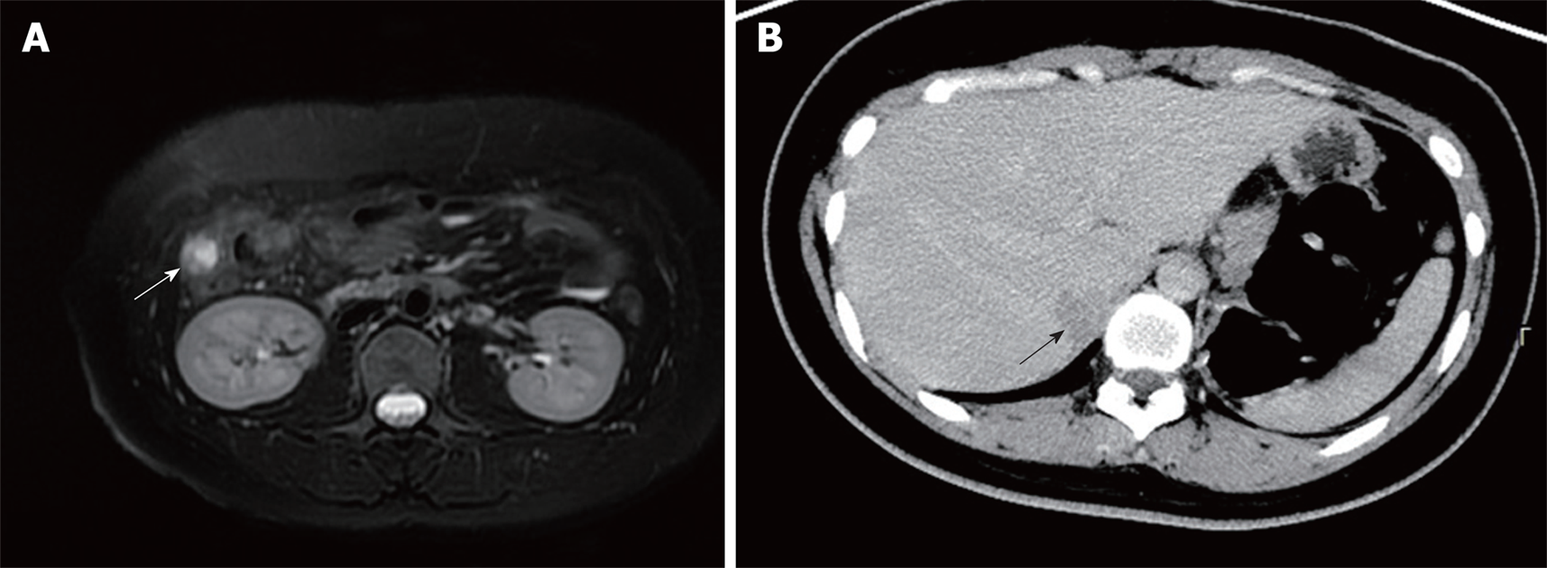
Figure 4 Computed tomography and magnetic resonance imaging at follow-up.
A: Abdominal magnetic resonance imaging (31 mo post-operatively) revealed a right paracolic nodule (white arrow); B: Abdominal enhanced computed tomography showed a low-density shadow in the liver (black arrow).
- Citation: Dai J, He HC, Huang X, Sun FK, Zhu Y, Xu DF. Long-term survival of a patient with a large adrenal primitive neuroectodermal tumor: A case report. World J Clin Cases 2019; 7(3): 340-346
- URL: https://www.wjgnet.com/2307-8960/full/v7/i3/340.htm
- DOI: https://dx.doi.org/10.12998/wjcc.v7.i3.340


