Copyright
©The Author(s) 2019.
World J Clin Cases. Dec 26, 2019; 7(24): 4321-4326
Published online Dec 26, 2019. doi: 10.12998/wjcc.v7.i24.4321
Published online Dec 26, 2019. doi: 10.12998/wjcc.v7.i24.4321
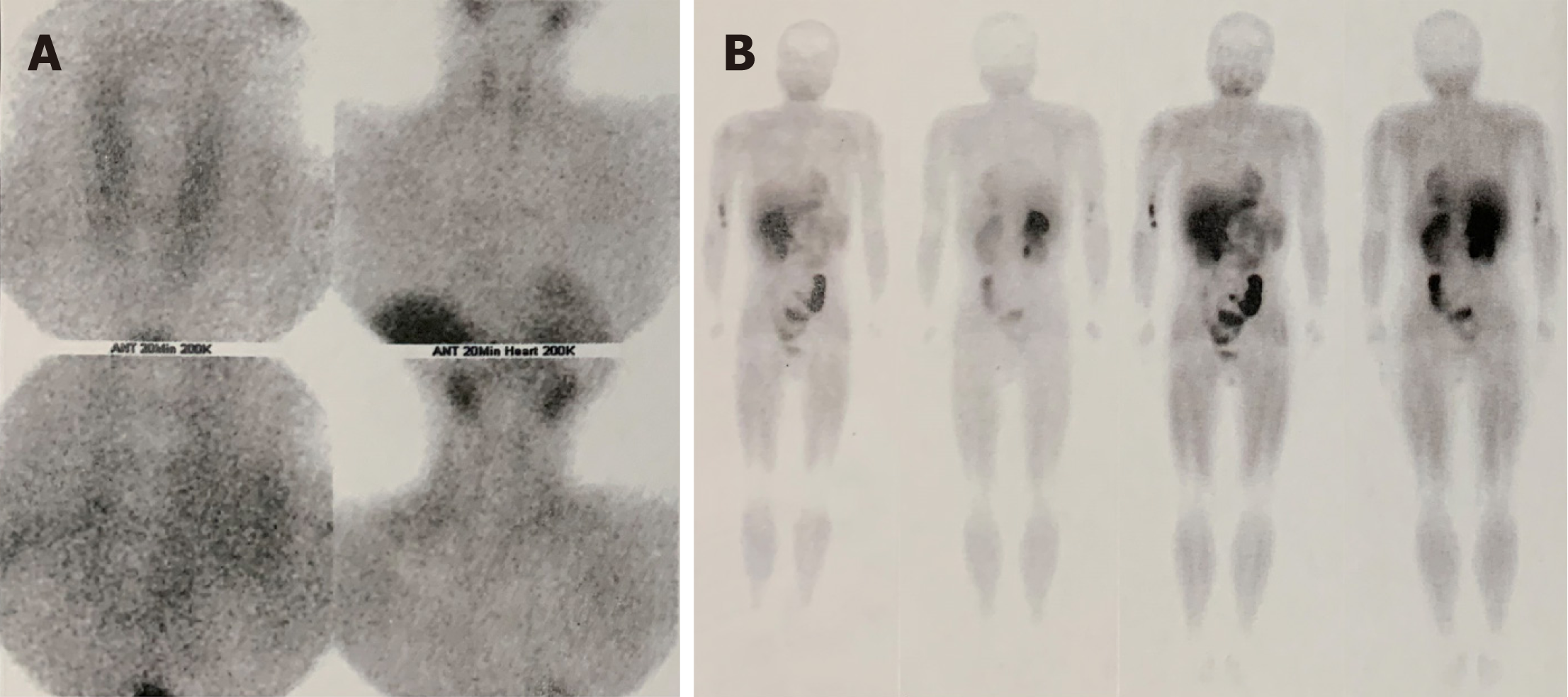
Figure 1 Ultrasound of parathyroid revealed no abnormal changes in the region of parathyroids, nor in 99mTc-methoxyisobutyl isonitrile imaging.
A-B showed that there was no abnormality of parathyroid in 99mTc-methoxyisobutyl isonitrile imaging, nor any other abnormal radioactivity concentrations.
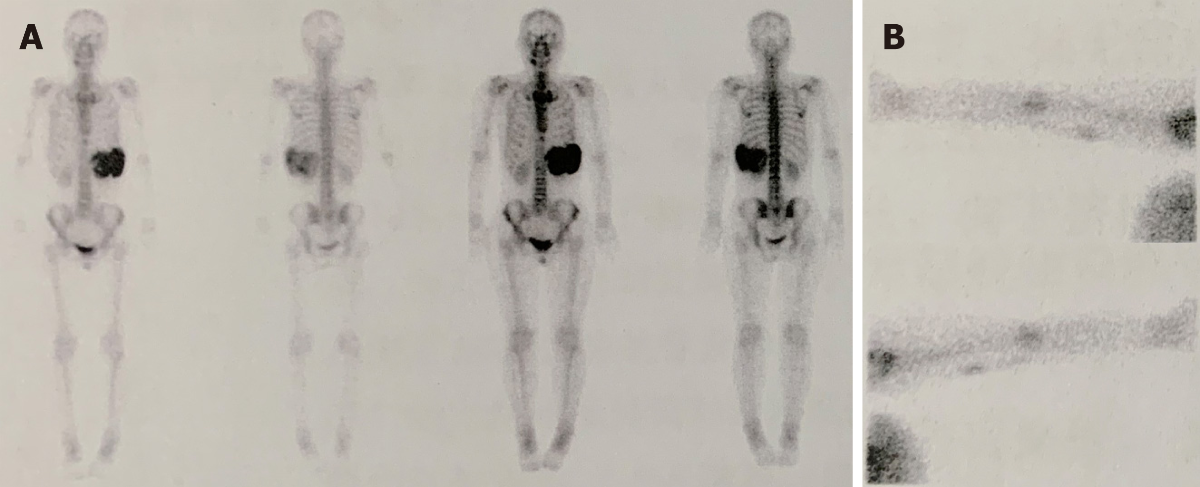
Figure 2 The whole-body bone imaging of the patient.
A: There was abnormal radioactivity concentrations at the position of the left 7-9th ribs; B: The tracer extravasation was shown in the right arm.
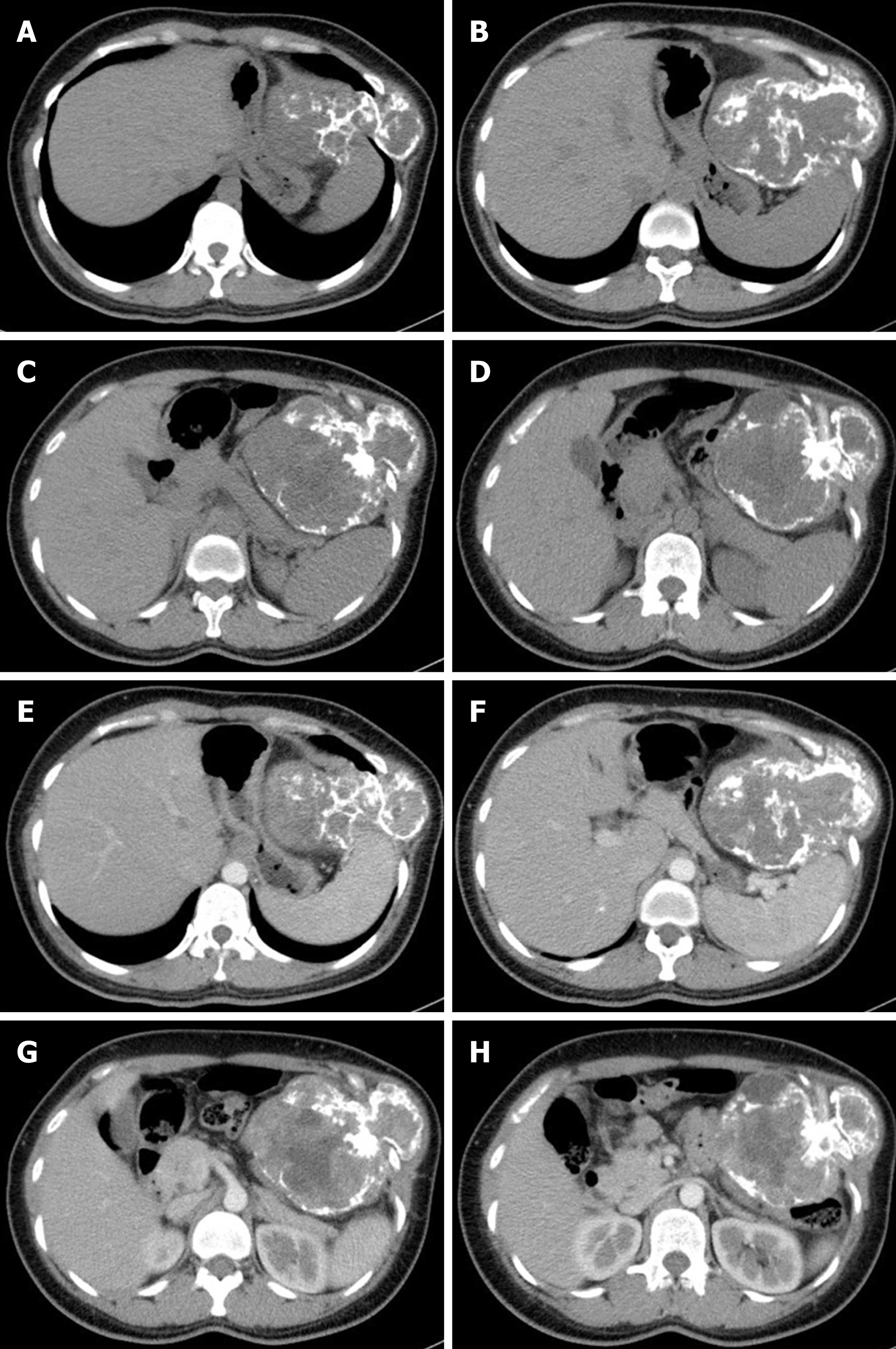
Figure 3 Enhanced chest and abdominal computed tomography revealed the rib mass.
A-D: Plain scan; E-H: Enhanced scan.
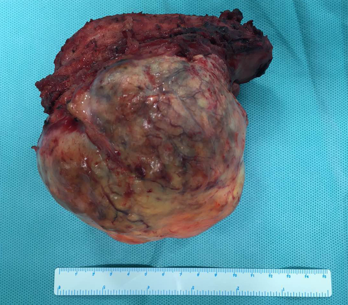
Figure 4 Gross pathology of the mass originating from the 9-10th ribs.
- Citation: Wang WD, Zhang N, Qu Q, He XD. Huge brown tumor of the rib in an unlocatable hyperparathyroidism patient with “self-recovered” serum calcium and parathyroid hormone: A case report. World J Clin Cases 2019; 7(24): 4321-4326
- URL: https://www.wjgnet.com/2307-8960/full/v7/i24/4321.htm
- DOI: https://dx.doi.org/10.12998/wjcc.v7.i24.4321


