Copyright
©The Author(s) 2019.
World J Clin Cases. Dec 26, 2019; 7(24): 4314-4320
Published online Dec 26, 2019. doi: 10.12998/wjcc.v7.i24.4314
Published online Dec 26, 2019. doi: 10.12998/wjcc.v7.i24.4314
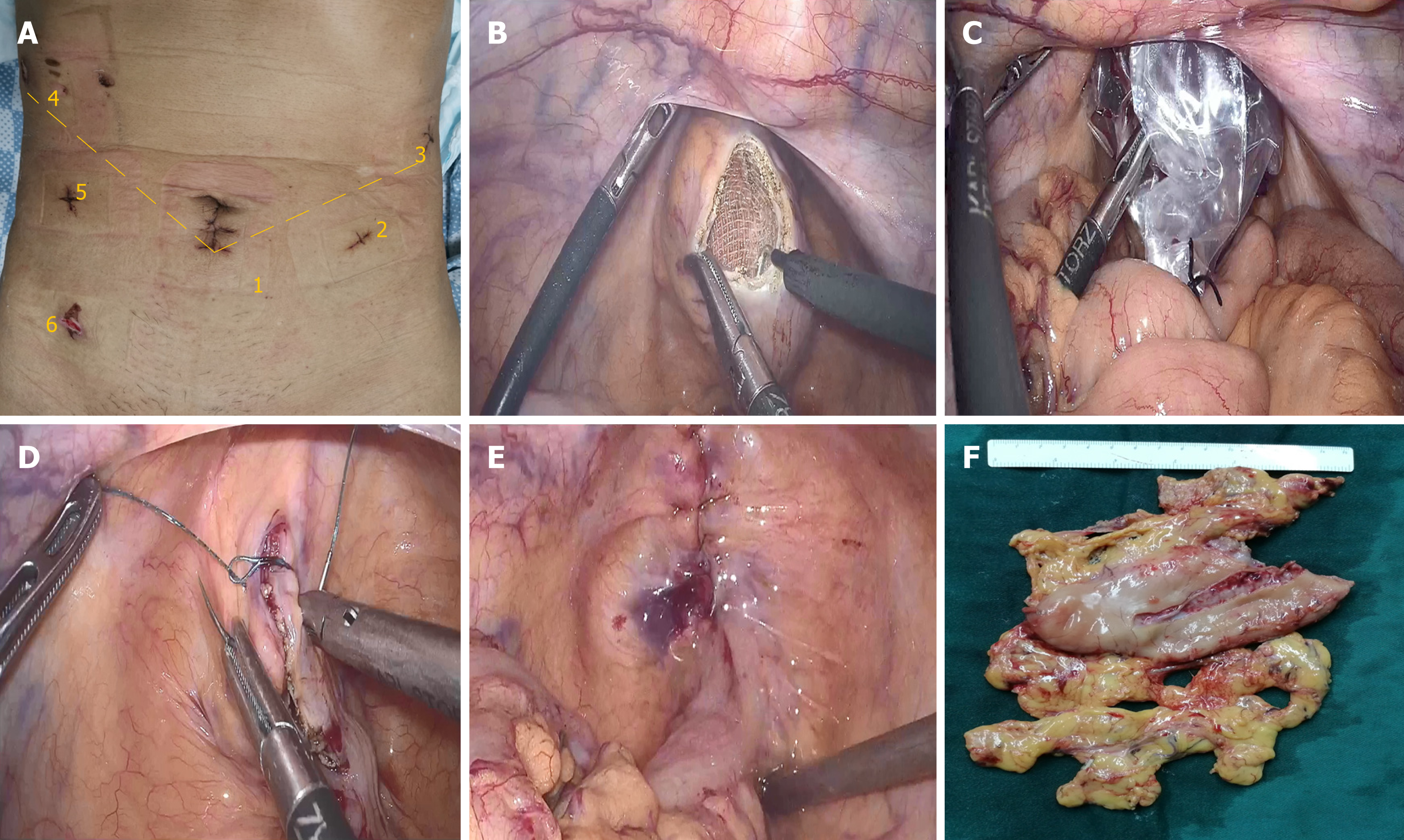
Figure 1 One more trocar was placed into the right lower abdomen to assist with specimen extraction.
A: Position of the trocars; B: The anterior wall of the upper rectum was cut using electrocautery; C: The specimen bag was gently pulled out of the body; D: Barbed wire sutures were used to close the incision; E: The rectal incision was completely closed; F: The specimen of distal gastric cancer.
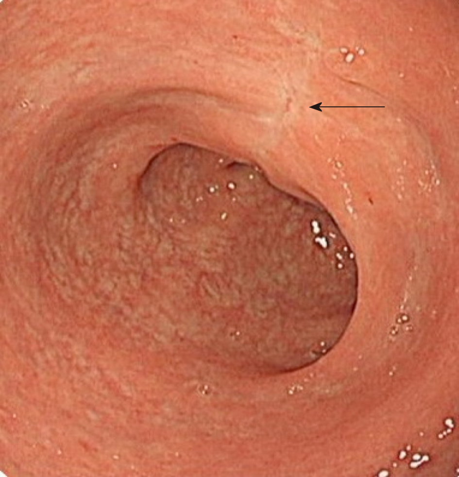
Figure 2 Colonoscopy showed no stenosis in the rectal cavity at 3 mo after surgery (arrow).
- Citation: Sun P, Wang XS, Liu Q, Luan YS, Tian YT. Natural orifice specimen extraction with laparoscopic radical gastrectomy for distal gastric cancer: A case report. World J Clin Cases 2019; 7(24): 4314-4320
- URL: https://www.wjgnet.com/2307-8960/full/v7/i24/4314.htm
- DOI: https://dx.doi.org/10.12998/wjcc.v7.i24.4314


