Copyright
©The Author(s) 2019.
World J Clin Cases. Dec 26, 2019; 7(24): 4292-4298
Published online Dec 26, 2019. doi: 10.12998/wjcc.v7.i24.4292
Published online Dec 26, 2019. doi: 10.12998/wjcc.v7.i24.4292
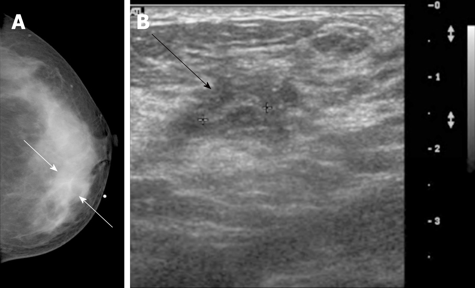
Figure 1 Breast mammography and ultrasonography findings.
A: Mammography (craniocaudal view) shows an asymmetry at the palpable site of the left inner breast (white arrows); B: Ultrasonography shows a tubular structure inside, approximately 1 cm long and with a circular diameter of 0.2 cm (black arrow).
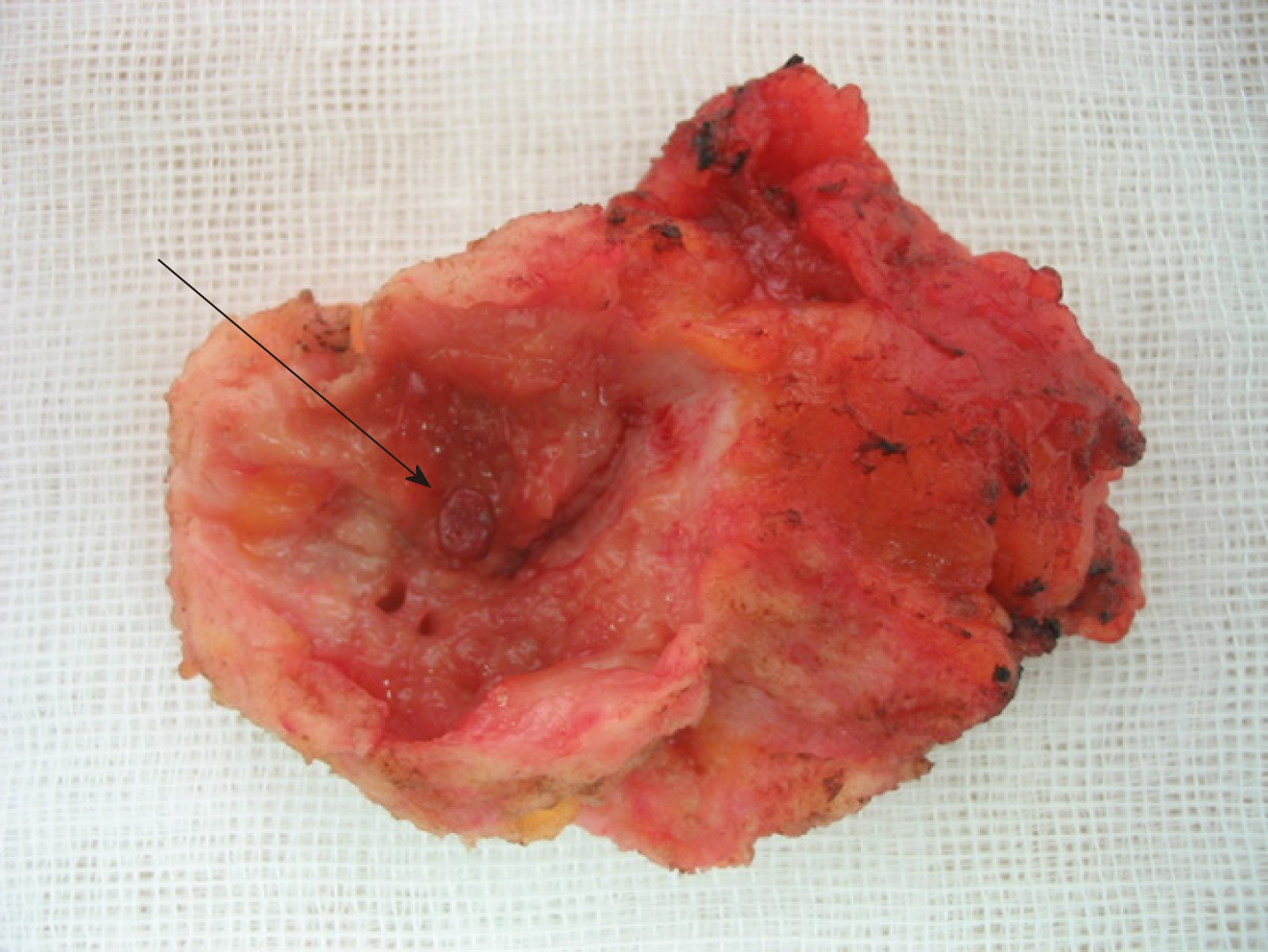
Figure 2 Gross specimen.
A cystic lesion of the excised soft tissue mass, with an irregular inner wall, was about 2.3 cm in longest diameter, and grayish white in color. The parasite was oval-shaped, red in color, and measured to be about 5 mm in longest diameter (arrow).
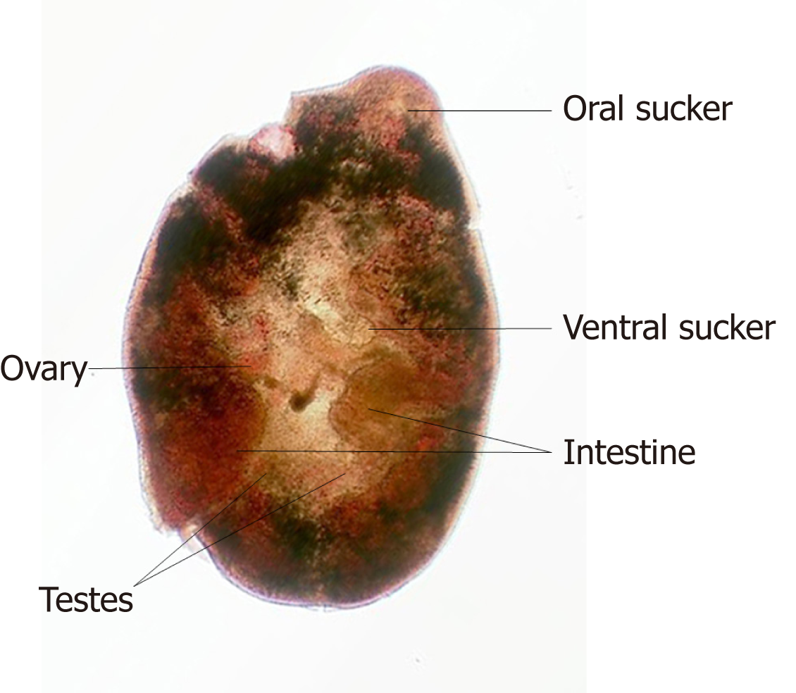
Figure 3 Paragonimus westermani juvenile worm.
The oral sucker is located on one end of the worm, and the ventral sucker is located at its center. The ovary and testes are stained red, and are less finely branched compared to that of adult worms. The intestines of the worm take a brown color and occupy the lateral fields.
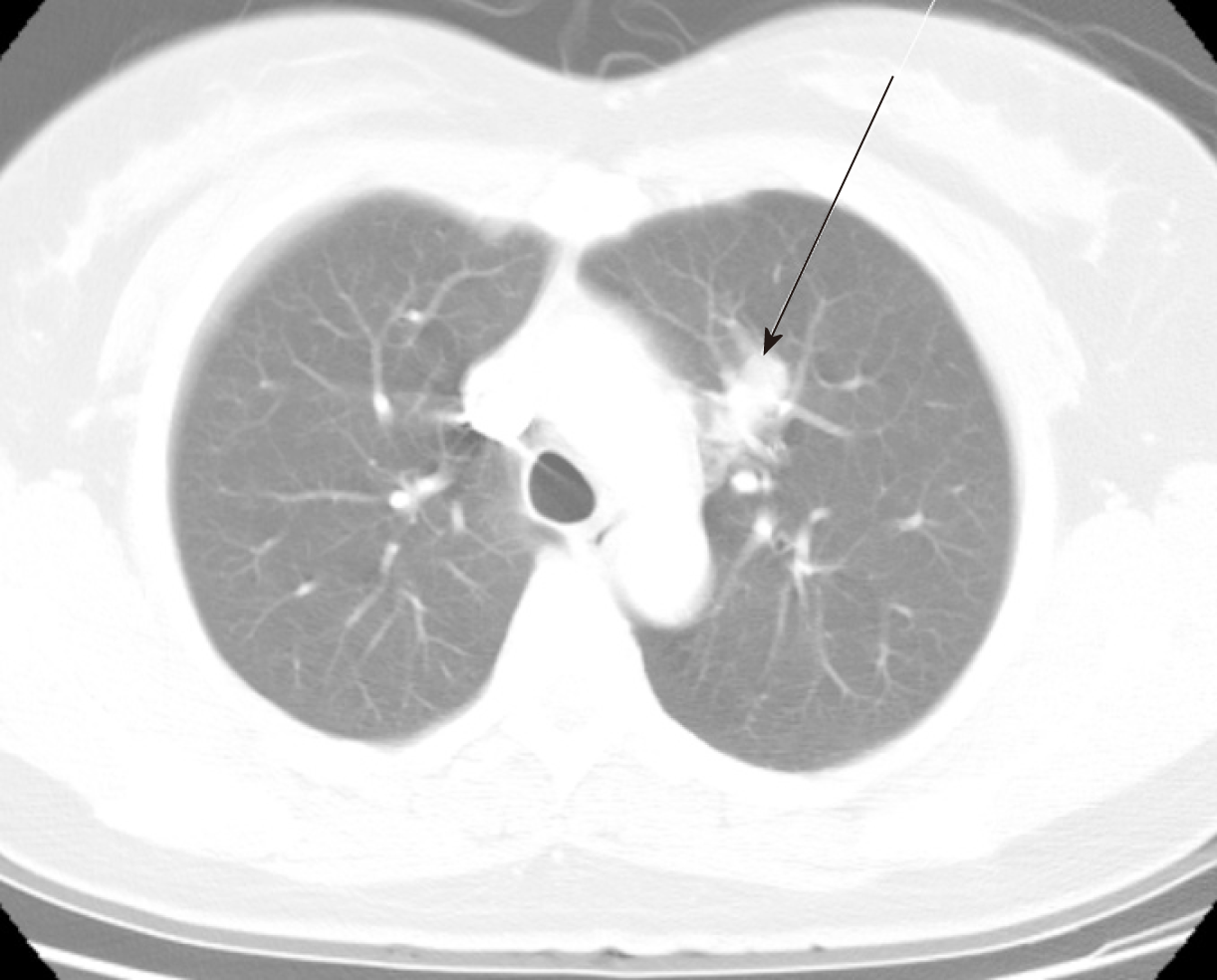
Figure 4 Chest computed tomography findings.
18 mm sized elongated nodule at left upper lobe of the lung (arrow).
- Citation: Oh MY, Chu A, Park JH, Lee JY, Roh EY, Chai YJ, Hwang KT. Simultaneous Paragonimus infection involving the breast and lung: A case report. World J Clin Cases 2019; 7(24): 4292-4298
- URL: https://www.wjgnet.com/2307-8960/full/v7/i24/4292.htm
- DOI: https://dx.doi.org/10.12998/wjcc.v7.i24.4292


