Copyright
©The Author(s) 2019.
World J Clin Cases. Dec 26, 2019; 7(24): 4196-4207
Published online Dec 26, 2019. doi: 10.12998/wjcc.v7.i24.4196
Published online Dec 26, 2019. doi: 10.12998/wjcc.v7.i24.4196
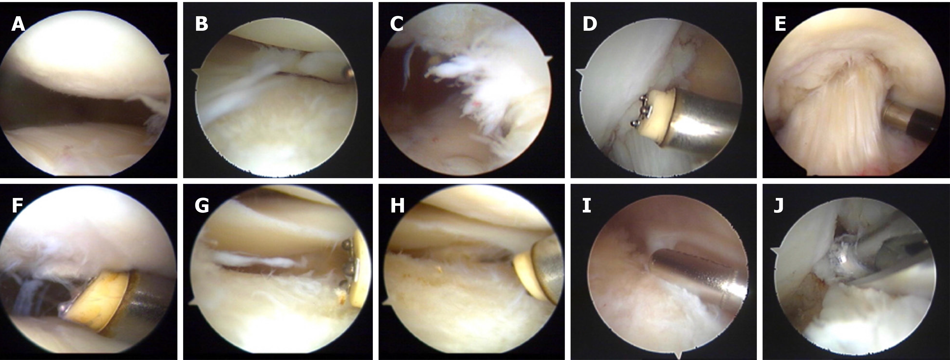
Figure 1 Surgery procedure and observations under arthroscopy.
A: Mild injury of patellofemoral articular cartilage; B: II-degree injury of lateral tibial plateau cartilage and meniscus injury; C: Synovial hyperplasia; D: Intercondylar fossa notch and epiphyseal hyperplasia; E: Evaluation of the tension of the anterior cruciate ligament; F: Trimming the cartilage surface of the tibia; G: Trimming the cartilage injury of the lateral tibial plateau; H: Trimming the injury of the lateral meniscus edge; I: Cleaning the synovial folds; J: Grinding the intercondylar fossa.
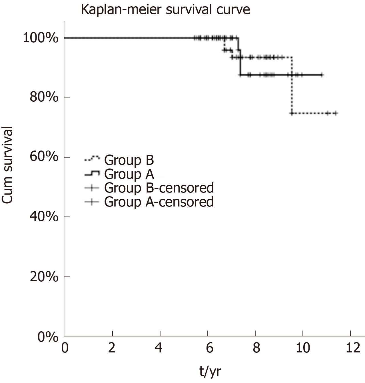
Figure 2 Twelve-year survivorship of patients between the two groups.
At the last follow-up, three patients in group A received revision with a survivorship of 95%, and four in group B received revision with a survivorship of 93.1%. The outcome showed no statistically significant difference for unicompartmental osteoarthritis patients receiving the two measures (P = 0.933).
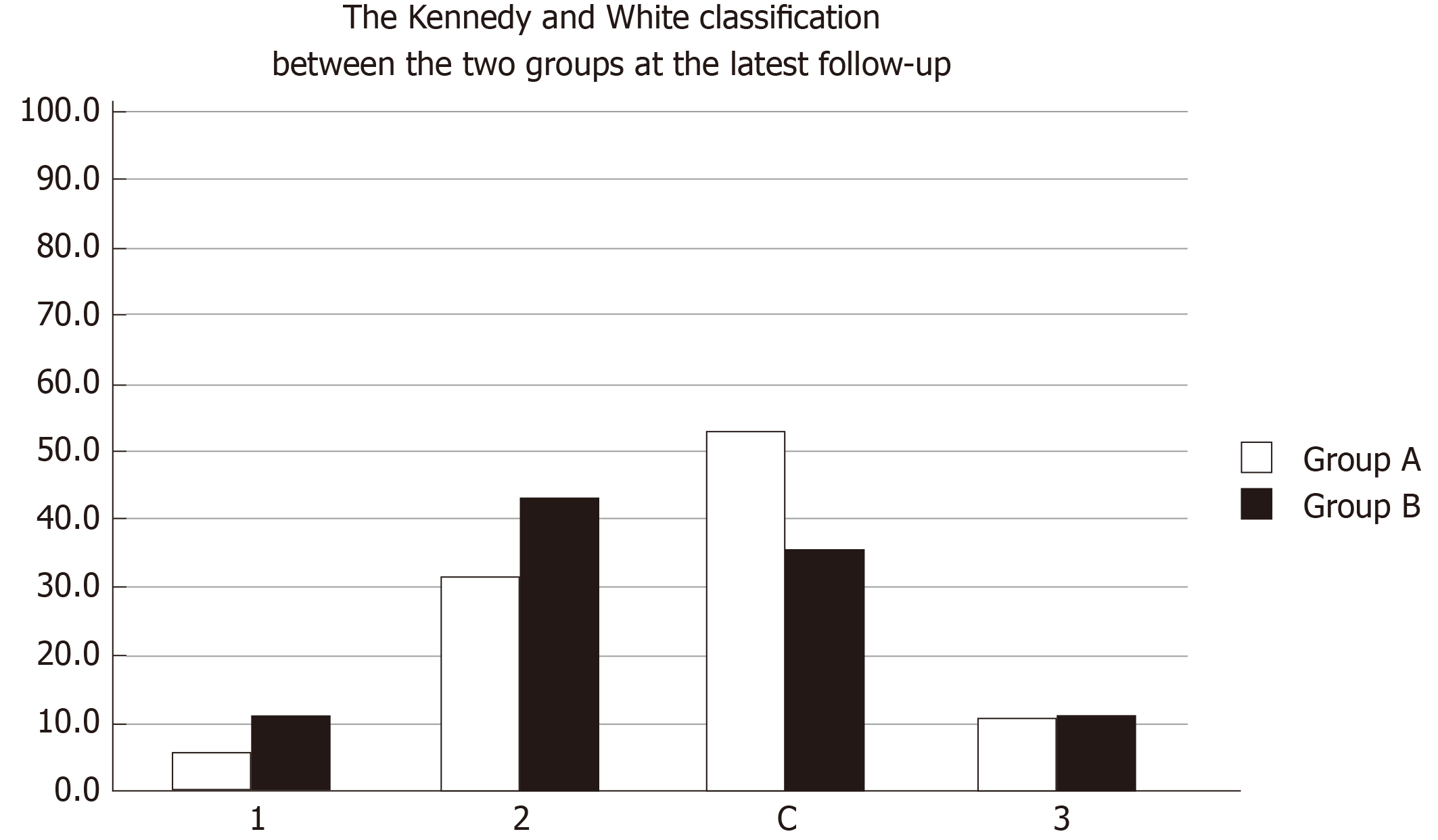
Figure 3 Kennedy and White classification between the two groups.
We defined valgus in area 3, varus in area 0 or 1, and alignment correction in area 2 or C.
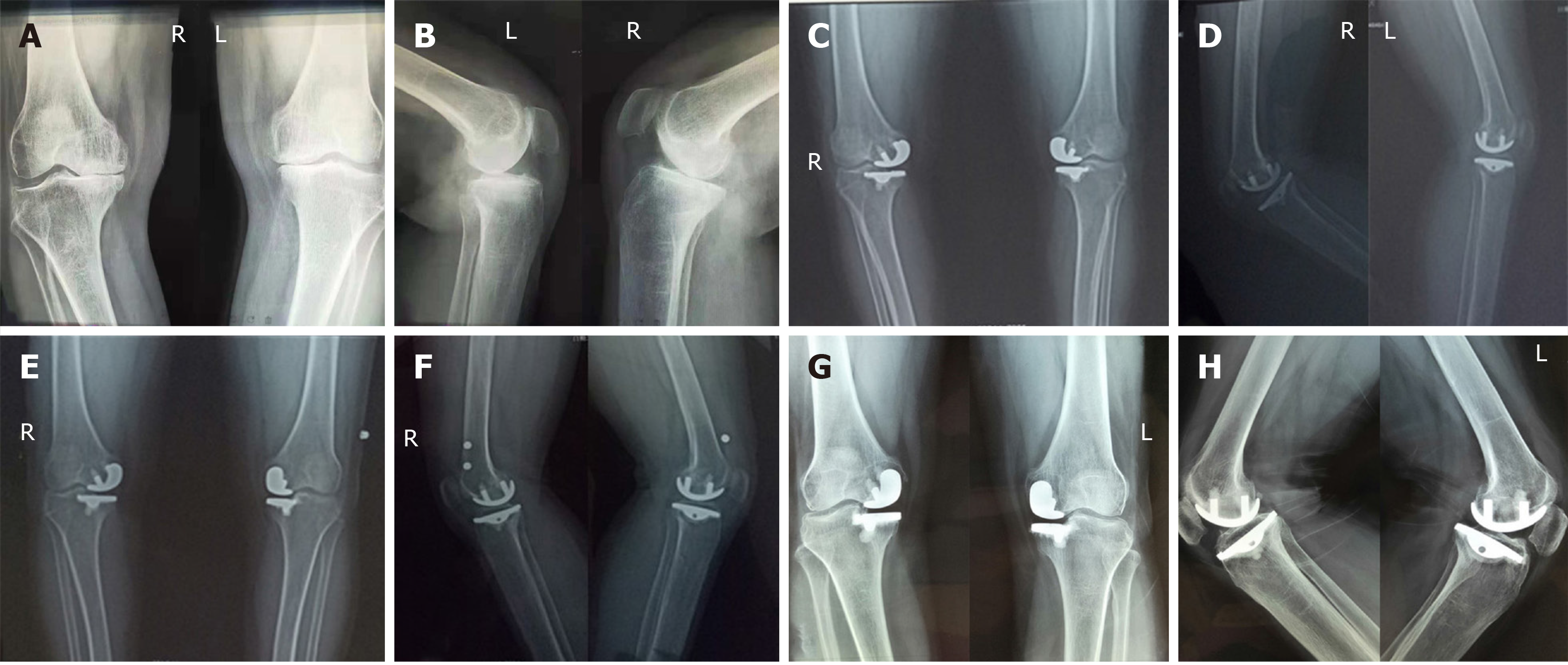
Figure 4 Images of a 60-year-old woman.
a and b: Positive lateral radiographs of the knee joint showing severe stenosis in the medial compartment of the knee. The patient's symptoms were pain in the medial knee and the knee joint flexed to 140°. Unicompartmental knee arthritis was performed on both knees and the postoperative recovery was good without complications. She was discharged from the hospital after 18 d of hospitalization; C and D: Positive lateral radiographs of the knee joints taken at 3 mo after the operation; E and F: Positive lateral radiographs of the knee joints taken at 6 months after the operation; G and H: Positive lateral radiographs of the knee joints taken at 7 years after the operation. The X-rays showed partial loosening of the prosthesis. The main symptom of the patient at present was pain in the medial side of the knee joint when standing or walking, and the symptoms were not significantly improved before the operation.
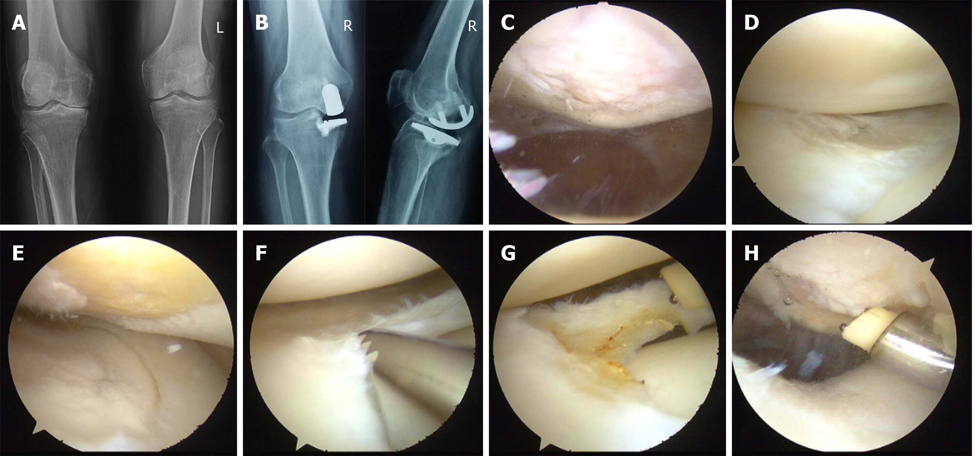
Figure 5 Surgical procedure and images of a 53-year-old woman.
A: Preoperative radiographs showing unicompartmental osteoarthritis on the right knee; B: Image taken at 8 years after arthroscopy combined with unicondylar knee arthroplasty showing that the prosthesis is in good position and the patient is experiencing good recovery as well as normal knee mobility. C: IV-degree injury of patellofemoral articular cartilage; D: Normal cartilage and meniscus injury of lateral compartment; E: IV-degree injury of internal femoral cartilage; F and G: Trimming the lateral meniscus injury site; H: Trimming the wound of patellofemoral articular cartilage.
- Citation: Wang HR, Li ZL, Li J, Wang YX, Zhao ZD, Li W. Arthroscopy combined with unicondylar knee arthroplasty for treatment of isolated unicompartmental knee arthritis: A long-term comparison. World J Clin Cases 2019; 7(24): 4196-4207
- URL: https://www.wjgnet.com/2307-8960/full/v7/i24/4196.htm
- DOI: https://dx.doi.org/10.12998/wjcc.v7.i24.4196


