Copyright
©The Author(s) 2019.
World J Clin Cases. Dec 6, 2019; 7(23): 4137-4143
Published online Dec 6, 2019. doi: 10.12998/wjcc.v7.i23.4137
Published online Dec 6, 2019. doi: 10.12998/wjcc.v7.i23.4137
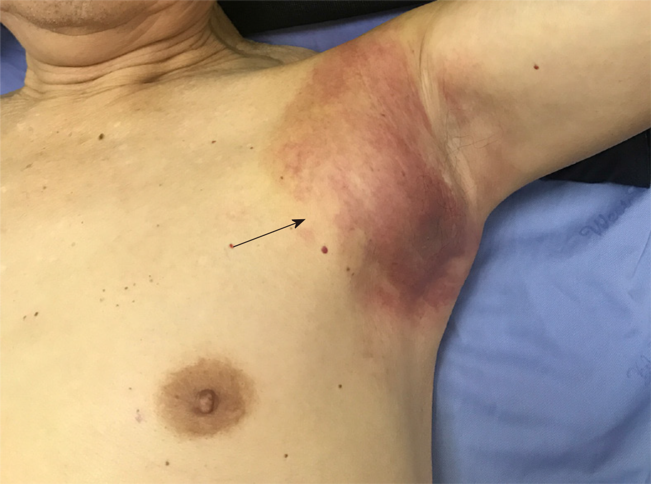
Figure 1 Left armpit subcutaneous metastasis arising from primary gastric cancer.
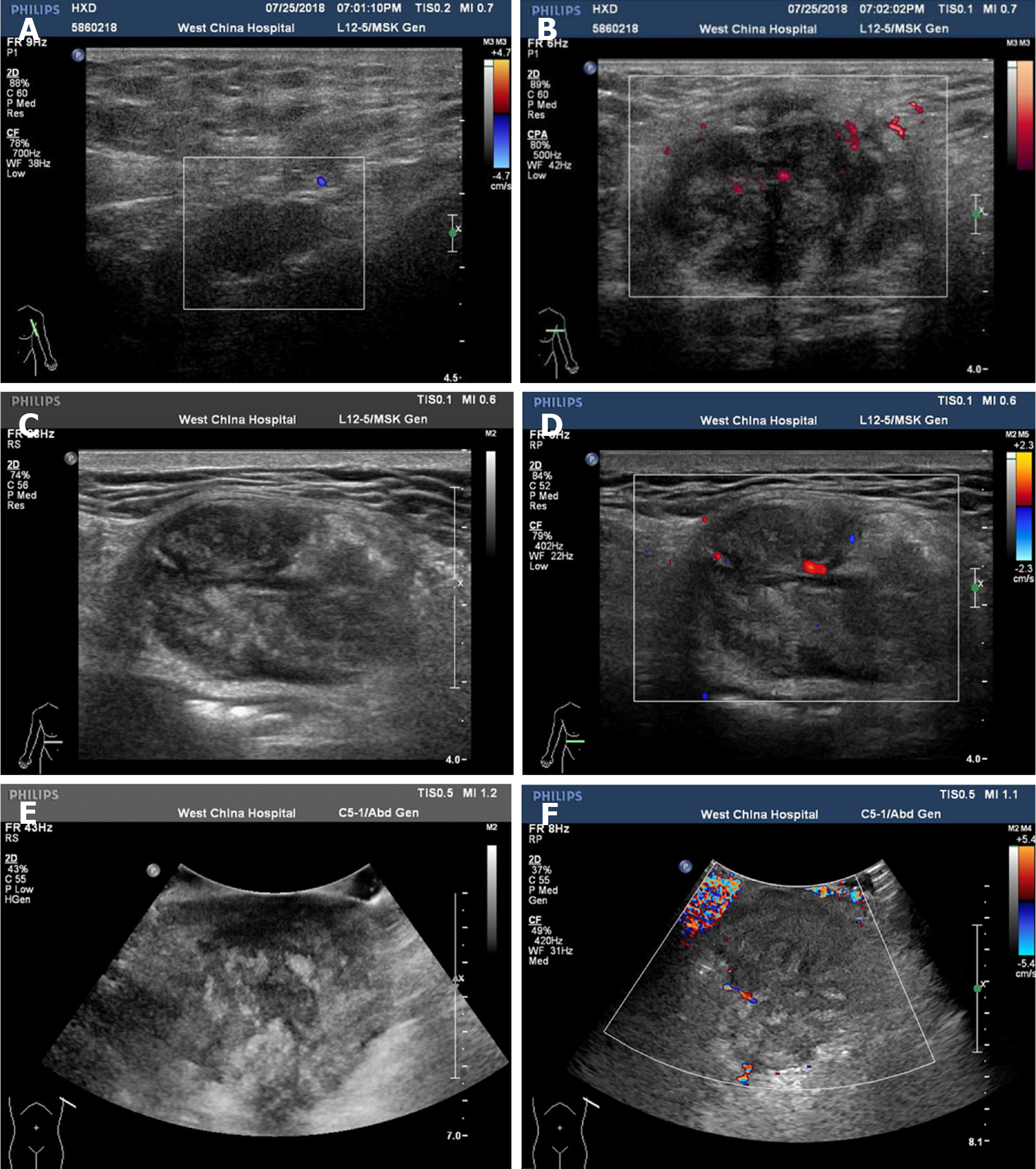
Figure 2 Color Doppler ultrasound images.
A and B: Initial color Doppler ultrasound images of the left axillary lump; C-F: Color Doppler ultrasound images of the left axillary lump after three months.
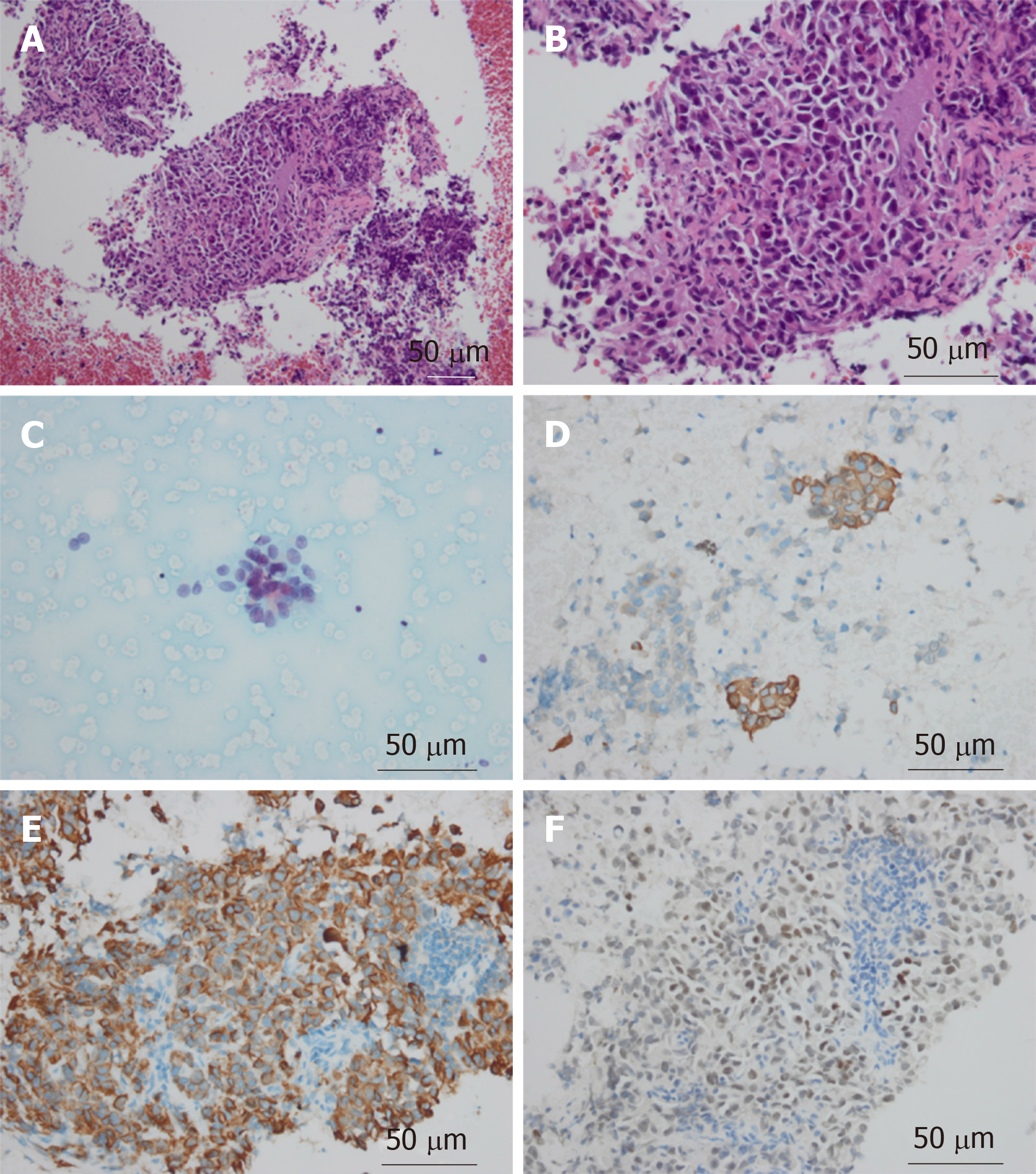
Figure 3 Pathological images of the axillary mass.
A-C: Stomach tumor cells were detected by cell smear (A: HE staining, ×200; B: HE staining, ×400; C: HE staining, ×400); D-F: The tumor cells were positive for CK20 (D), PCK (E), and CDX-2 (F) (immunohistochemical staining, ×400).
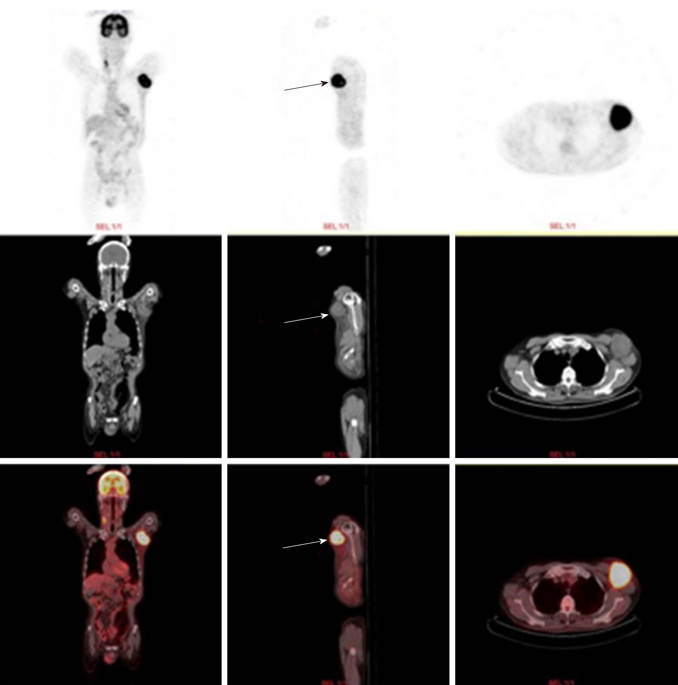
Figure 4 Positron emission tomography computed tomography examination showed that a 6.
2 cm × 5.5 cm soft tissue mass was visible in the left axilla (arrow).
- Citation: He FJ, Zhang P, Wang MJ, Chen Y, Zhuang W. Left armpit subcutaneous metastasis of gastric cancer: A case report. World J Clin Cases 2019; 7(23): 4137-4143
- URL: https://www.wjgnet.com/2307-8960/full/v7/i23/4137.htm
- DOI: https://dx.doi.org/10.12998/wjcc.v7.i23.4137


