Copyright
©The Author(s) 2019.
World J Clin Cases. Nov 26, 2019; 7(22): 3904-3911
Published online Nov 26, 2019. doi: 10.12998/wjcc.v7.i22.3904
Published online Nov 26, 2019. doi: 10.12998/wjcc.v7.i22.3904
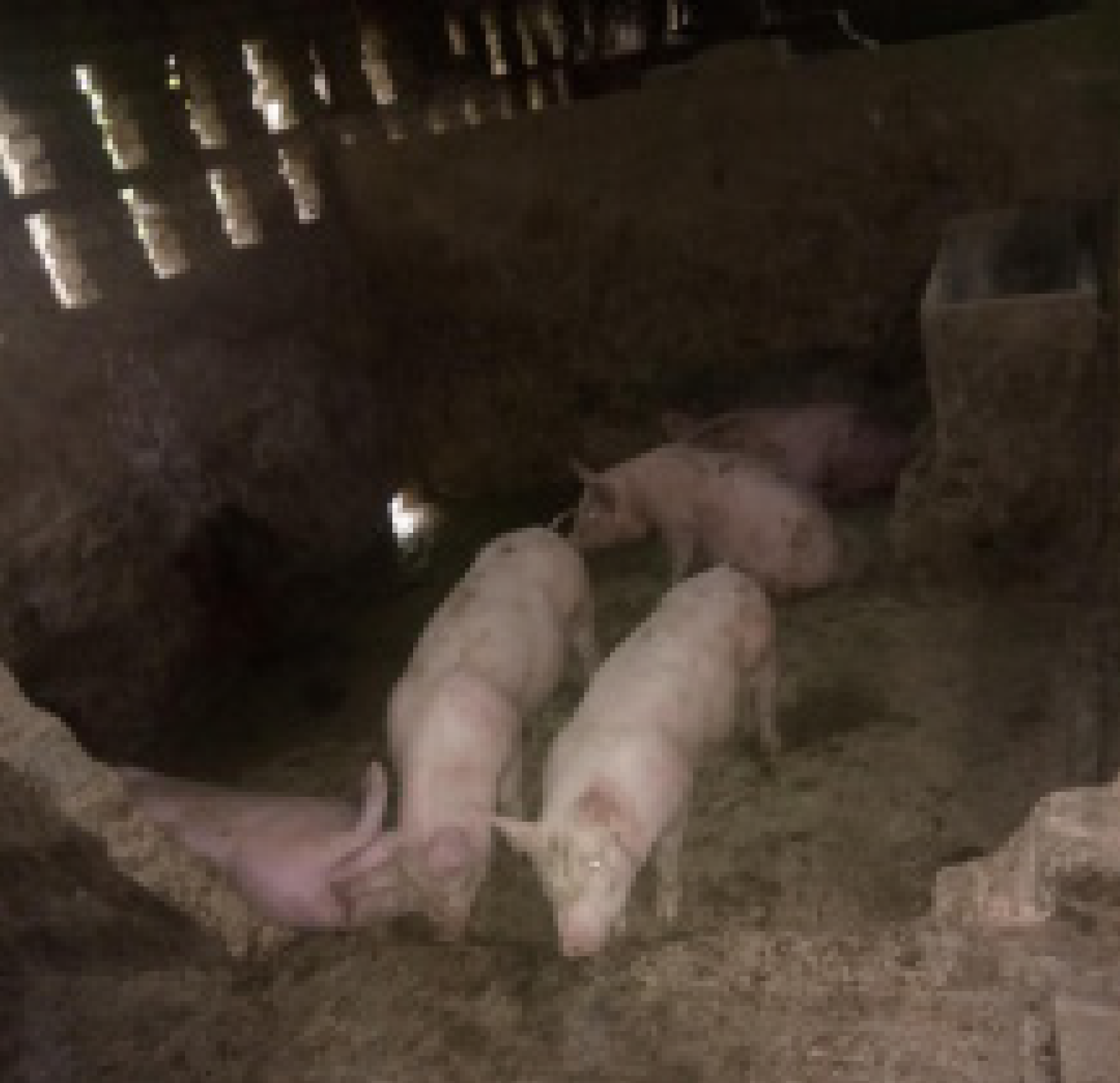
Figure 1 The pig farm environment.
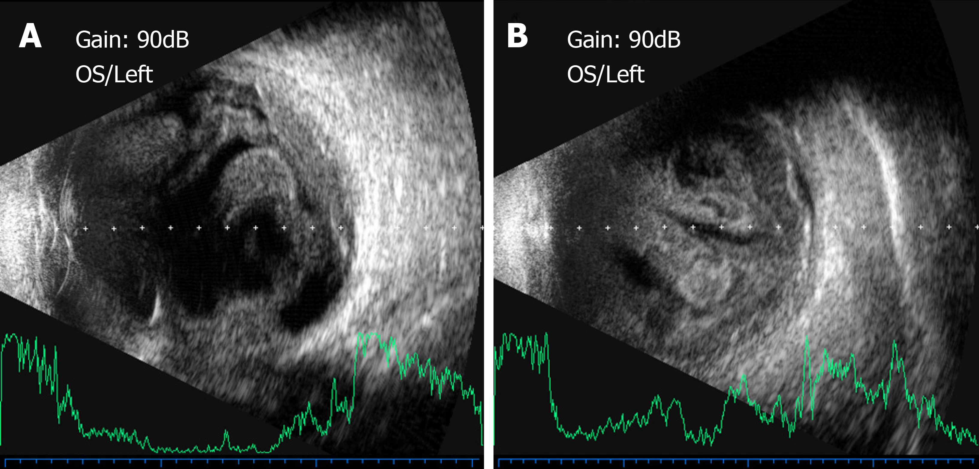
Figure 2 B-ultrasonography.
A: Vitreous opacities with retinal detachment; B: Deteriorating vitreous opacities and retinal detachment.
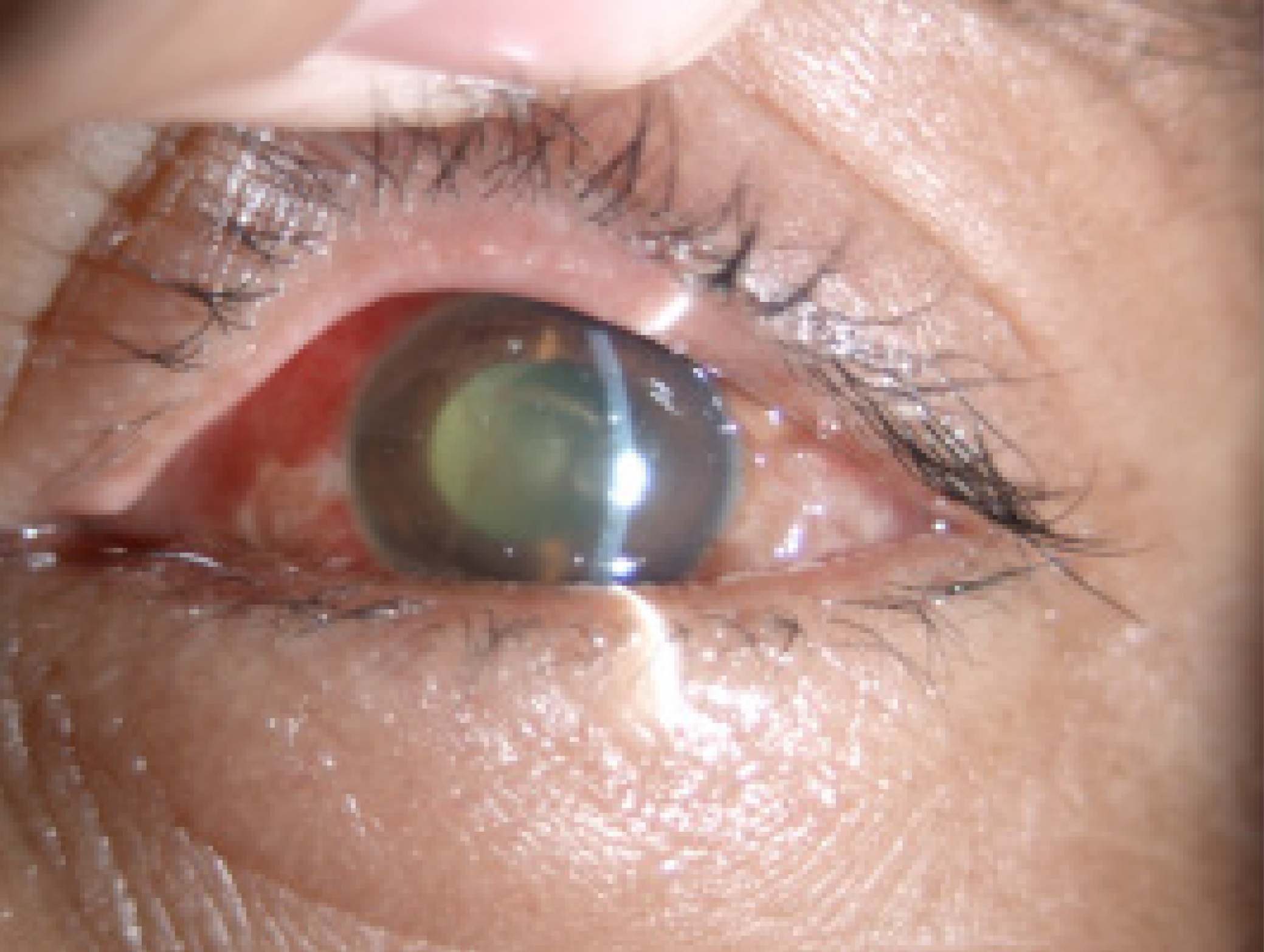
Figure 3 Slit-lamp examination showed reduced anterior segment inflammation.
- Citation: Bao QD, Liu TX, Xie M, Tian X. Exogenous endophthalmitis caused by Enterococcus casseliflavus: A case report. World J Clin Cases 2019; 7(22): 3904-3911
- URL: https://www.wjgnet.com/2307-8960/full/v7/i22/3904.htm
- DOI: https://dx.doi.org/10.12998/wjcc.v7.i22.3904


