Copyright
©The Author(s) 2019.
World J Clin Cases. Nov 26, 2019; 7(22): 3866-3871
Published online Nov 26, 2019. doi: 10.12998/wjcc.v7.i22.3866
Published online Nov 26, 2019. doi: 10.12998/wjcc.v7.i22.3866
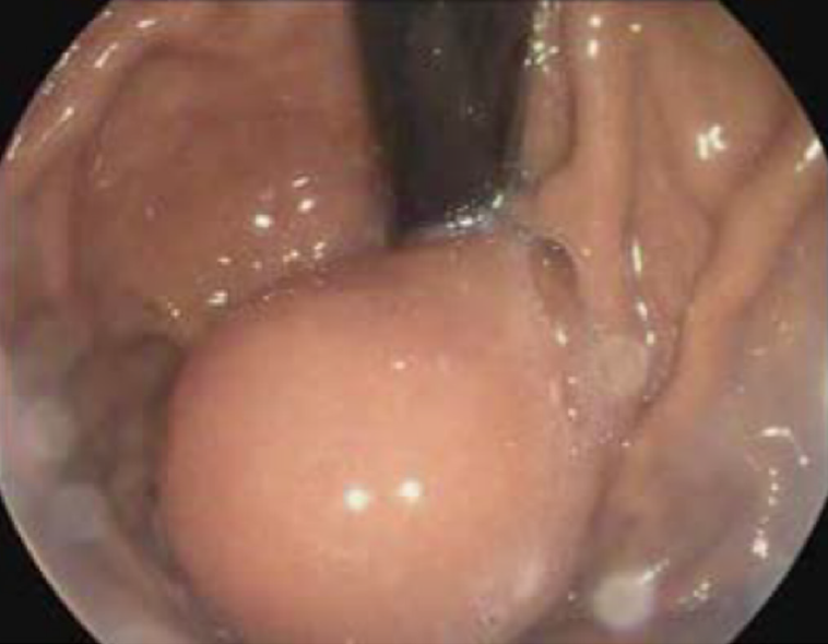
Figure 1 Upper endoscopy changes due to the gastric duplication cyst.
Figure 2 Endoscopic ultrasonography changes due to the gastric duplication cyst.
A: Endoscopic ultrasonography changes due to the gastric duplication cyst; B: Endoscopic ultrasonography-guided fine needle aspiration shows the gastric duplication cyst.
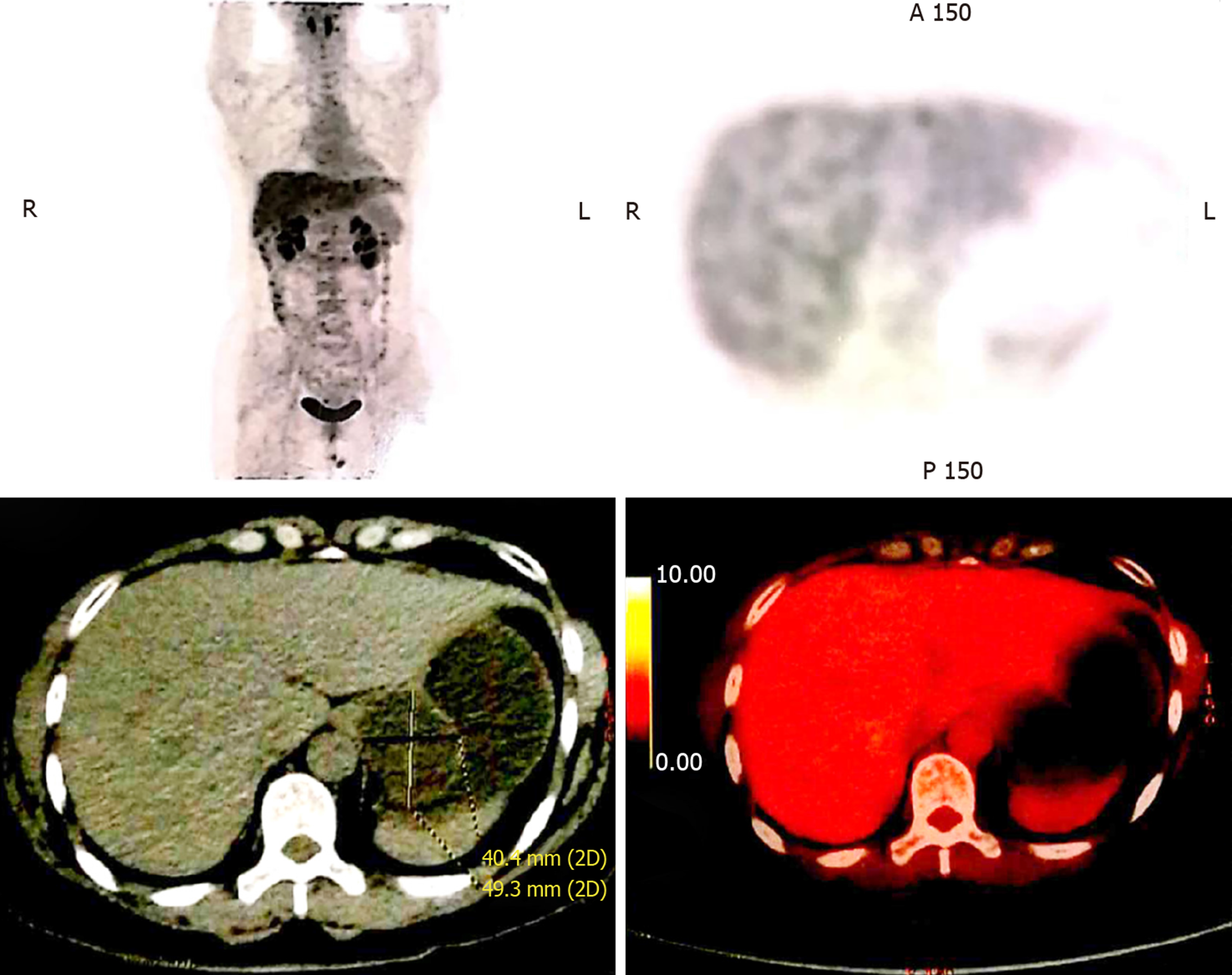
Figure 3 Positron emission tomography/computed tomography shows the gastric duplication cyst.
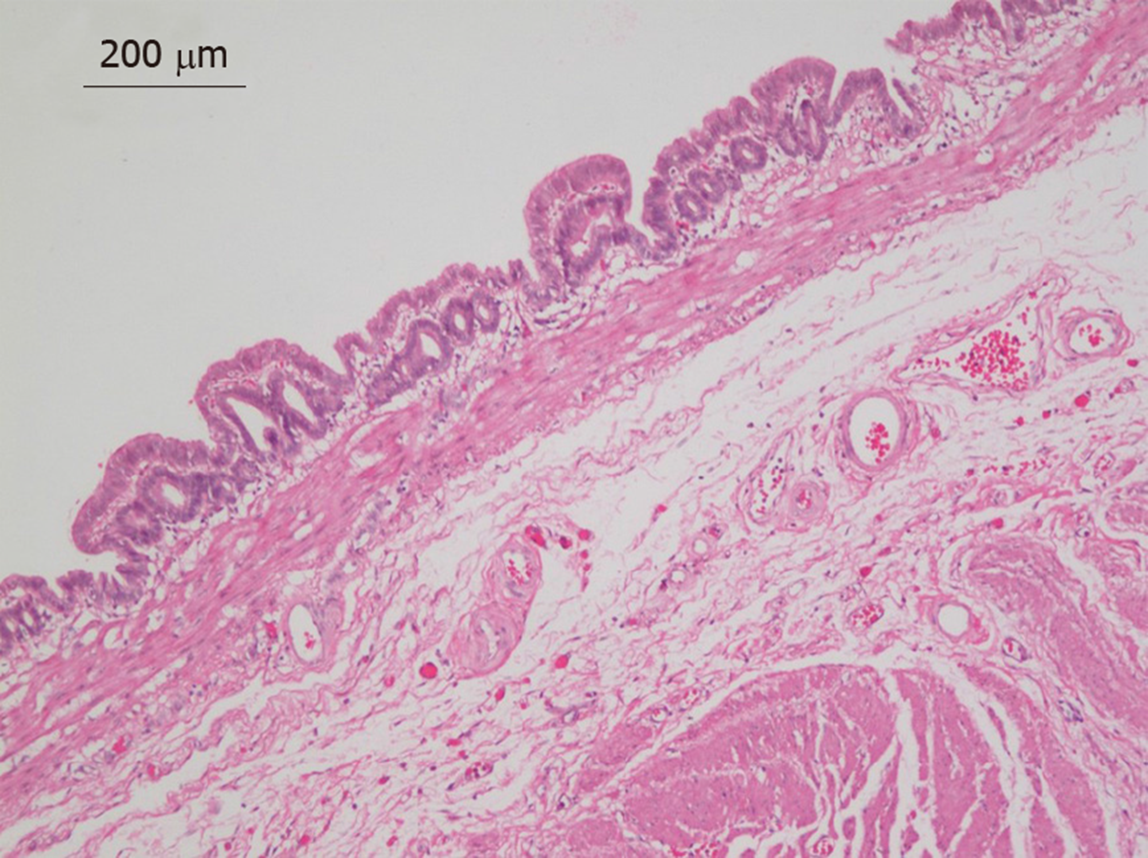
Figure 4 Pathological changes in the gastric duplication cyst (hematoxylin and eosin staining, ×100).
- Citation: Hu YB, Gui HW. Diagnosis of gastric duplication cyst by positron emission tomography/computed tomography: A case report. World J Clin Cases 2019; 7(22): 3866-3871
- URL: https://www.wjgnet.com/2307-8960/full/v7/i22/3866.htm
- DOI: https://dx.doi.org/10.12998/wjcc.v7.i22.3866


