Copyright
©The Author(s) 2019.
World J Clin Cases. Nov 26, 2019; 7(22): 3838-3843
Published online Nov 26, 2019. doi: 10.12998/wjcc.v7.i22.3838
Published online Nov 26, 2019. doi: 10.12998/wjcc.v7.i22.3838
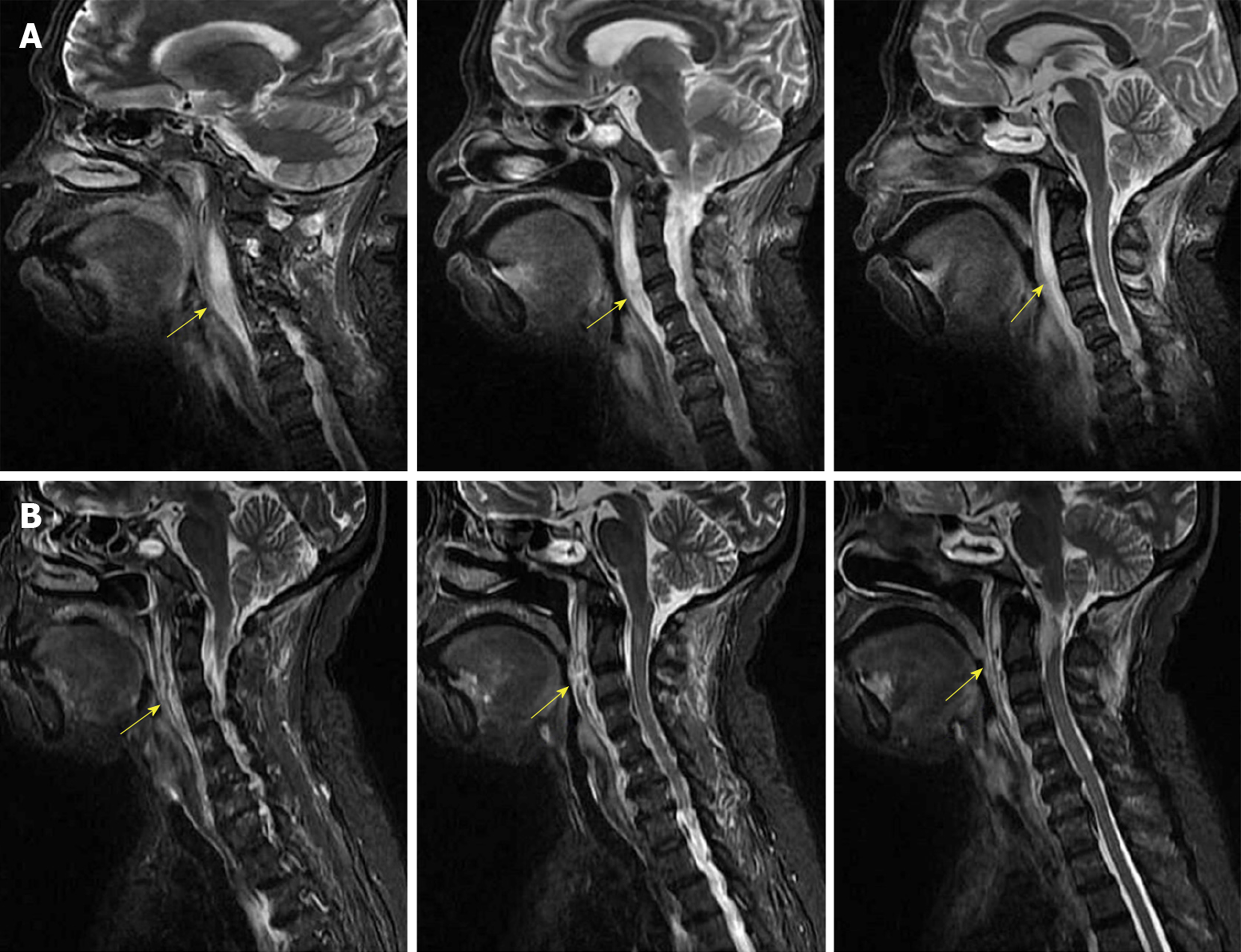
Figure 1 MRI findings.
A: Contrast-enhanced MRI shows the retropharyngeal abscess and obvious upper airway stenosis (yellow arrow); B: MRI suggests that upper airway stenosis was significantly alleviated after surgery (yellow arrow). MRI: Magnetic resonance imaging.
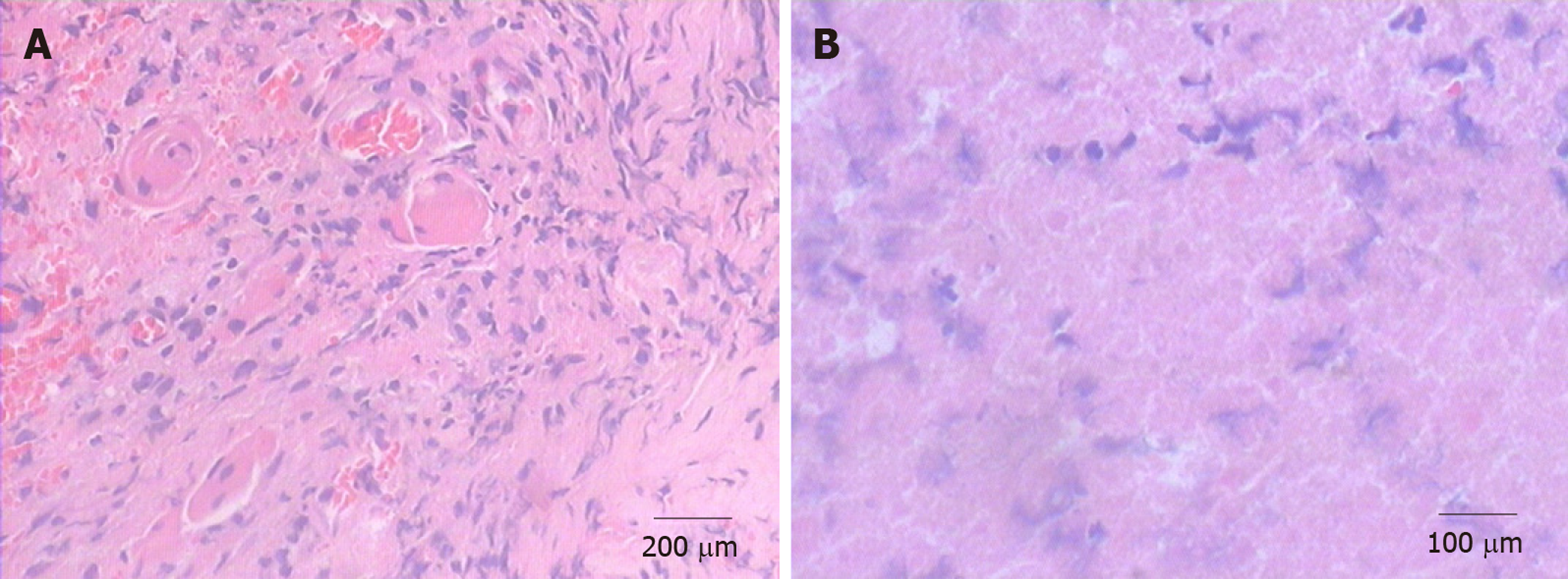
Figure 2 Histopathology findings.
A: Histopathology shows granulation hyperplasia (magnification × 200) in the retropharyngeal abscess tissues; B: Inflammatory necrosis (magnification × 400) in the retropharyngeal abscess tissues. HE: Hematoxylin and eosin.
- Citation: Lin J, Wu XM, Feng JX, Chen MF. Retropharyngeal abscess presenting as acute airway obstruction in a 66-year-old woman: A case report. World J Clin Cases 2019; 7(22): 3838-3843
- URL: https://www.wjgnet.com/2307-8960/full/v7/i22/3838.htm
- DOI: https://dx.doi.org/10.12998/wjcc.v7.i22.3838


