Copyright
©The Author(s) 2019.
World J Clin Cases. Nov 26, 2019; 7(22): 3812-3820
Published online Nov 26, 2019. doi: 10.12998/wjcc.v7.i22.3812
Published online Nov 26, 2019. doi: 10.12998/wjcc.v7.i22.3812
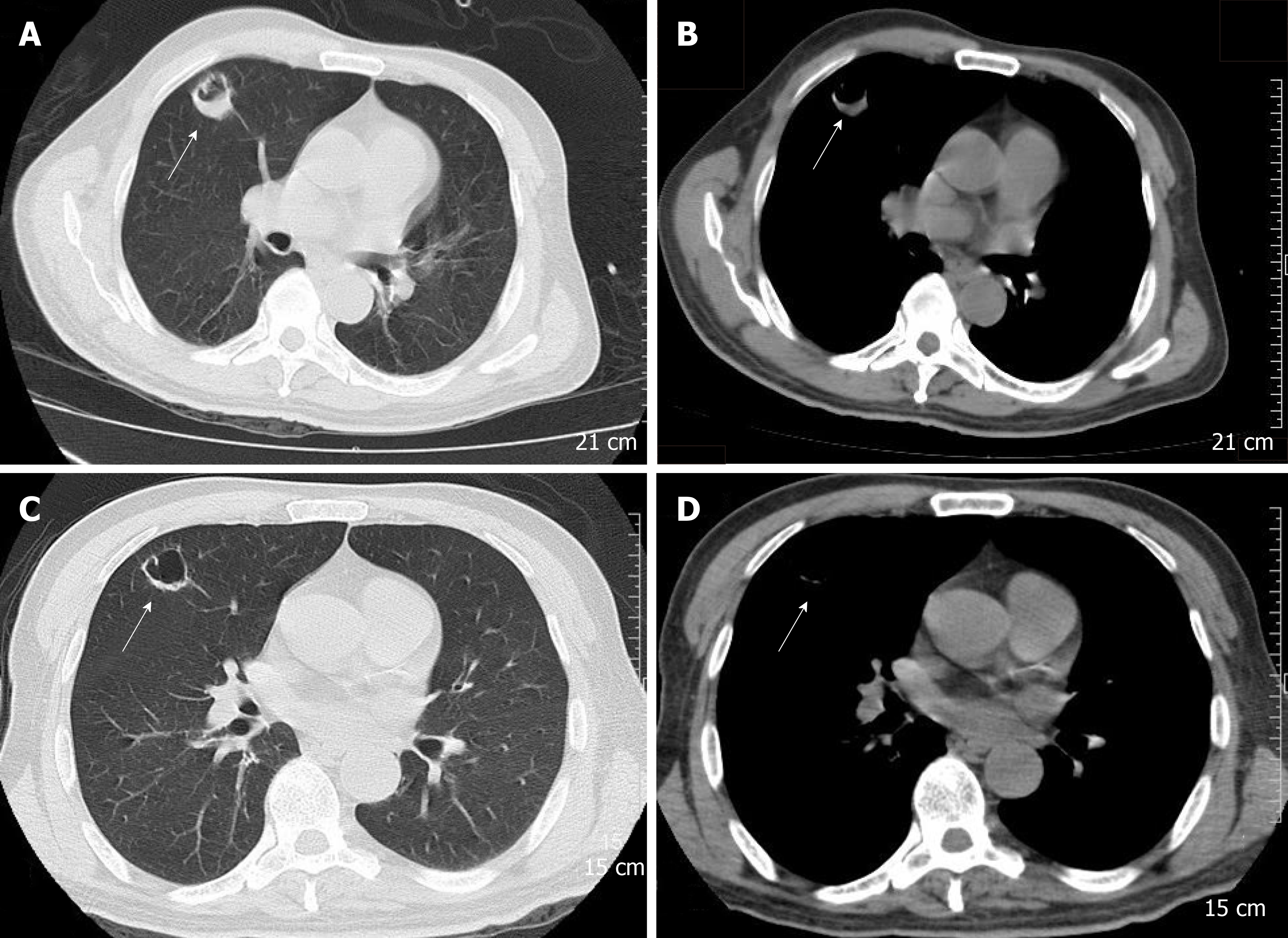
Figure 1 CT tomography scan at admission and 10 d later.
A, B: Axial CT images: lung window setting (A) and mediastinal window setting (B) (slice thickness 3 mm) showed a 25 mm × 27 mm, round, thick-walled cavity in the right upper lobe. C, D: Axial CT images: Lung window setting (C), and mediastinal window setting (D) (slice thickness 3 mm) showed that the thick-walled cavity had been absorbed to a 21 mm × 21 mm, thin-walled cavity after treatment for 10 d. CT: Computed tomography.
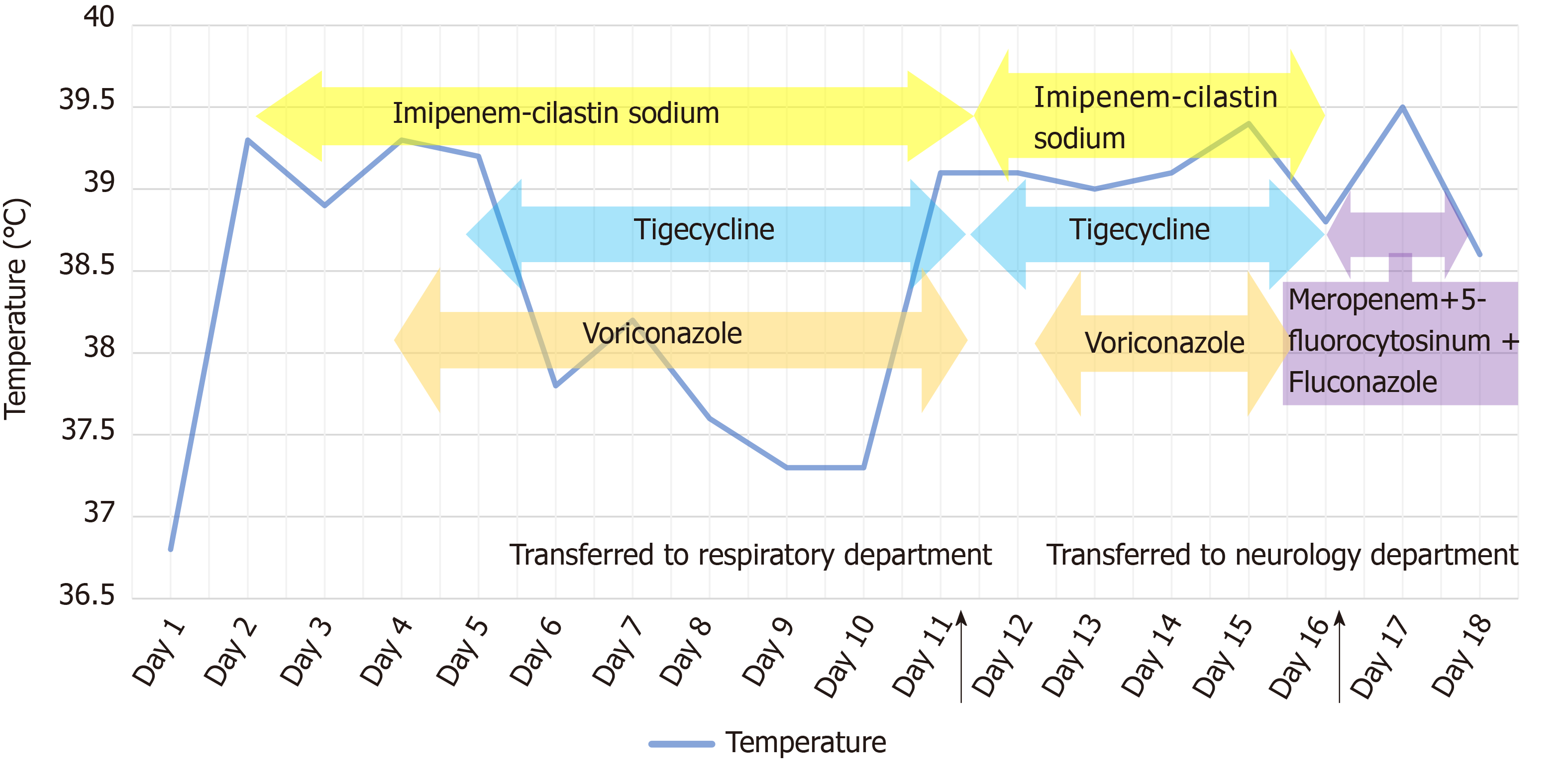
Figure 2 Body temperature and antibiotic use during hospitalization.
Antibiotic use: imipenem-cilastatin sodium 2 g every 8 hr (days 1-11) and 1 g every 6 hr (days 12-16), tigecycline 0.1 g every 12 hr (days 4-16), voriconazole 0.2 g every 12 hr (days 1-11 and 12-16), meropenem 2 g every 8 hr + 5-fluorocytosine 1.5 g every 6 hr + fluconazole 0.4 g every 12 hr (days 16-18).
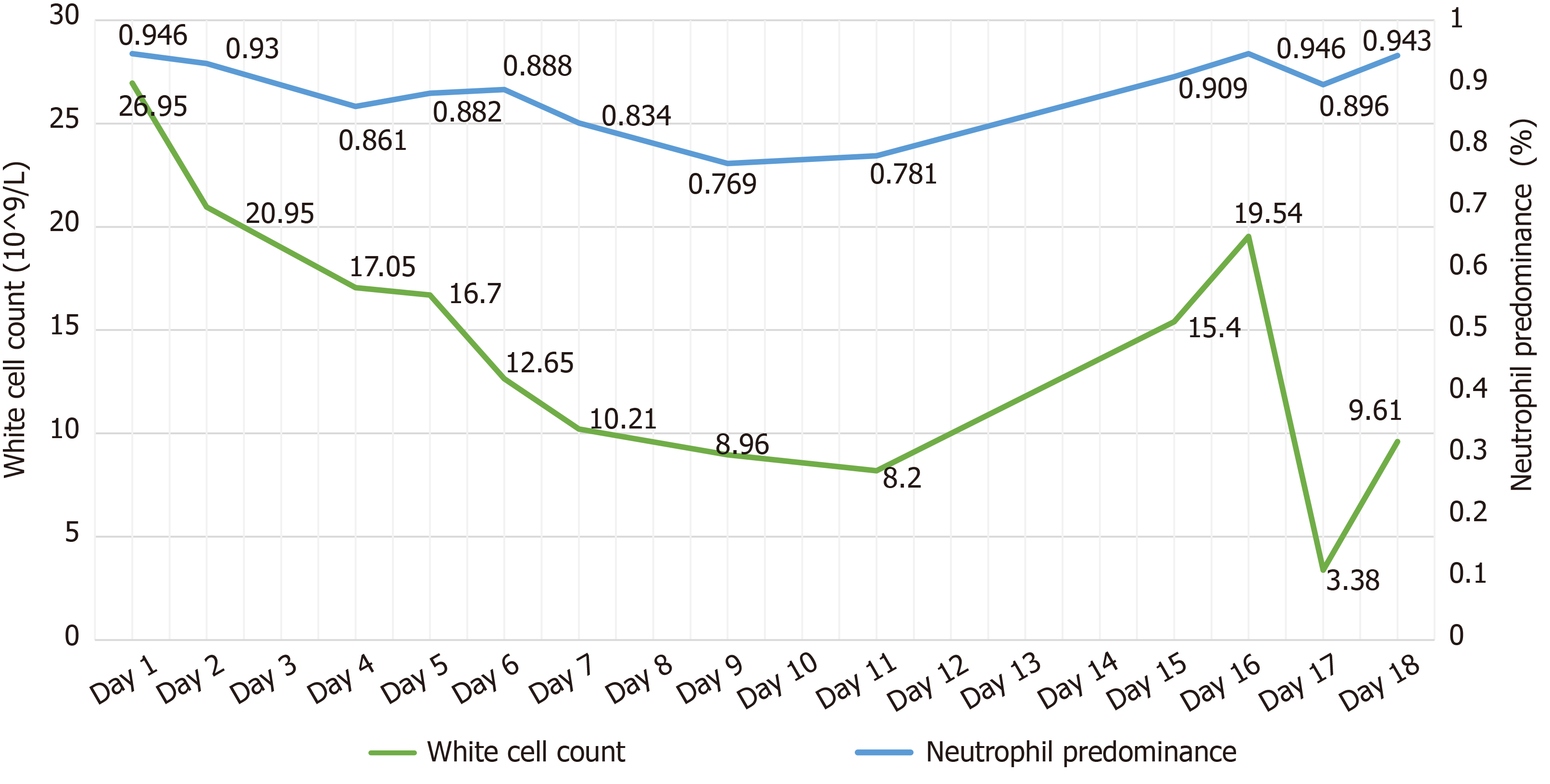
Figure 3 Change in white blood cell count and neutrophil predominance during hospitalization.
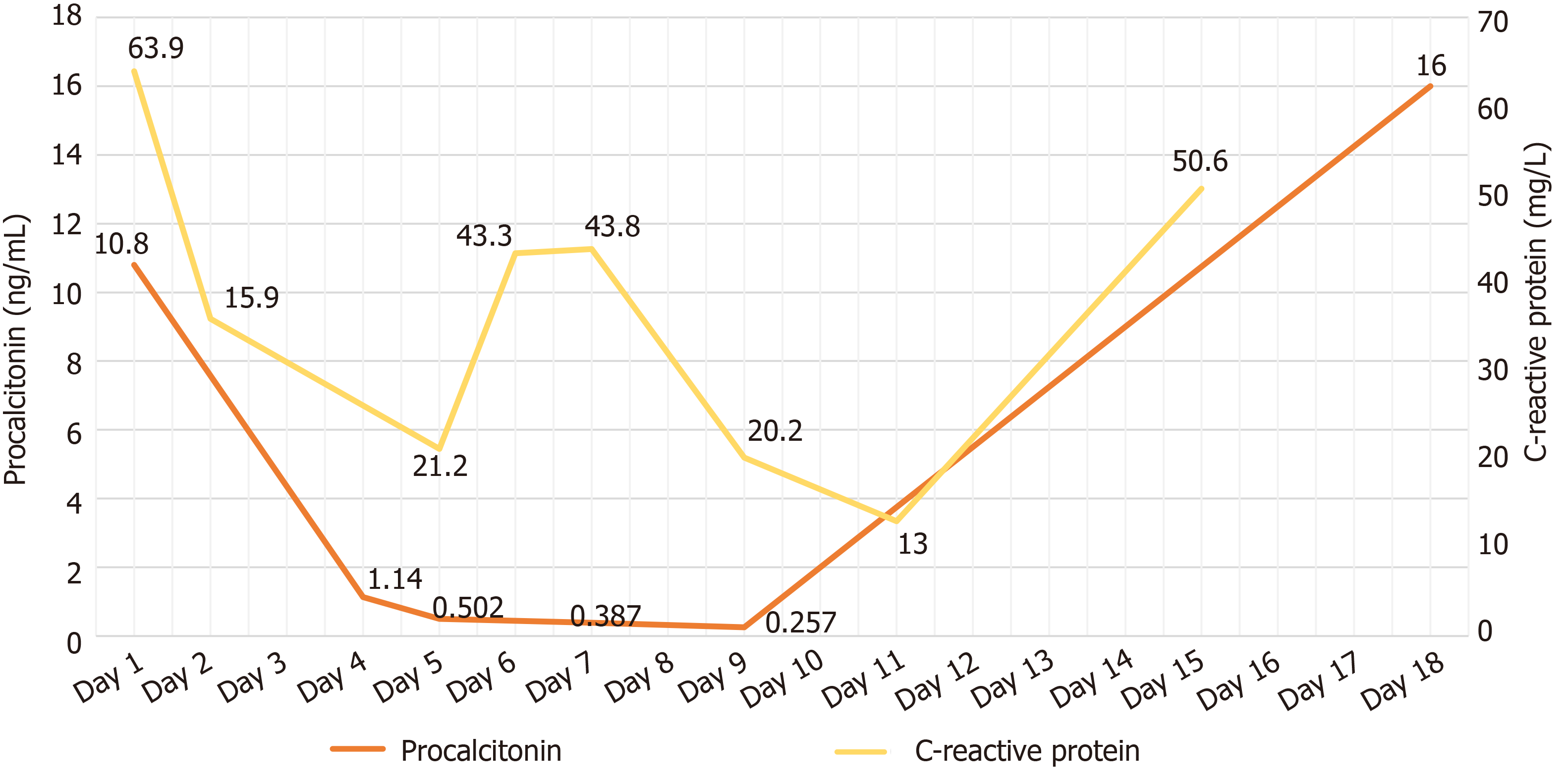
Figure 4 Change in C-creative protein and procalcitonin during hospitalization.
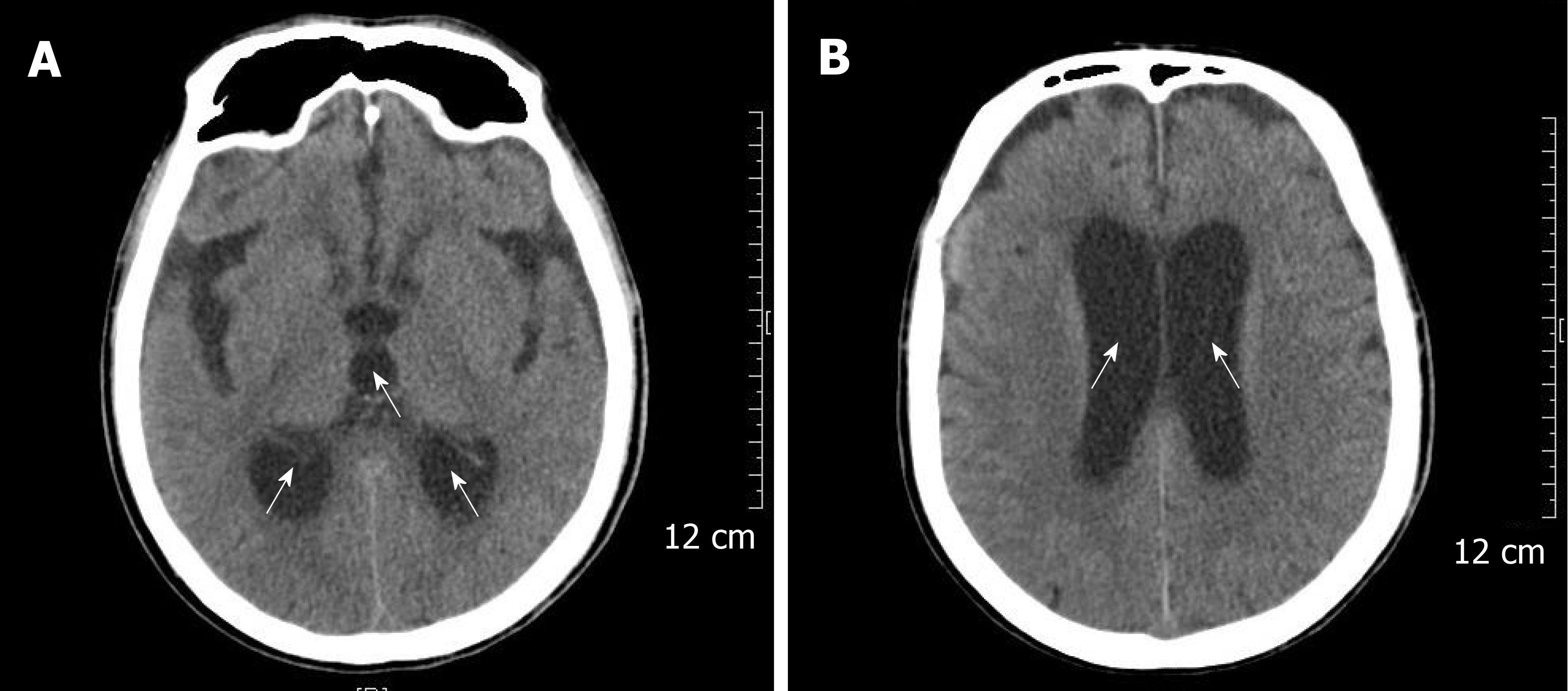
Figure 5 Axial unenhanced computed tomography scan of the head.
A: Axial unenhanced computed tomography scan of the head showed marked dilatation of the occipital horn of the lateral ventricles and third ventricles; B: Marked dilatation of the bilateral ventricle.
- Citation: Shi YF, Wang YK, Wang YH, Liu H, Shi XH, Li XJ, Wu BQ. Metastatic infection caused by hypervirulent Klebsiella pneumonia and co-infection with Cryptococcus meningitis: A case report. World J Clin Cases 2019; 7(22): 3812-3820
- URL: https://www.wjgnet.com/2307-8960/full/v7/i22/3812.htm
- DOI: https://dx.doi.org/10.12998/wjcc.v7.i22.3812


