Copyright
©The Author(s) 2019.
World J Clin Cases. Nov 26, 2019; 7(22): 3765-3771
Published online Nov 26, 2019. doi: 10.12998/wjcc.v7.i22.3765
Published online Nov 26, 2019. doi: 10.12998/wjcc.v7.i22.3765
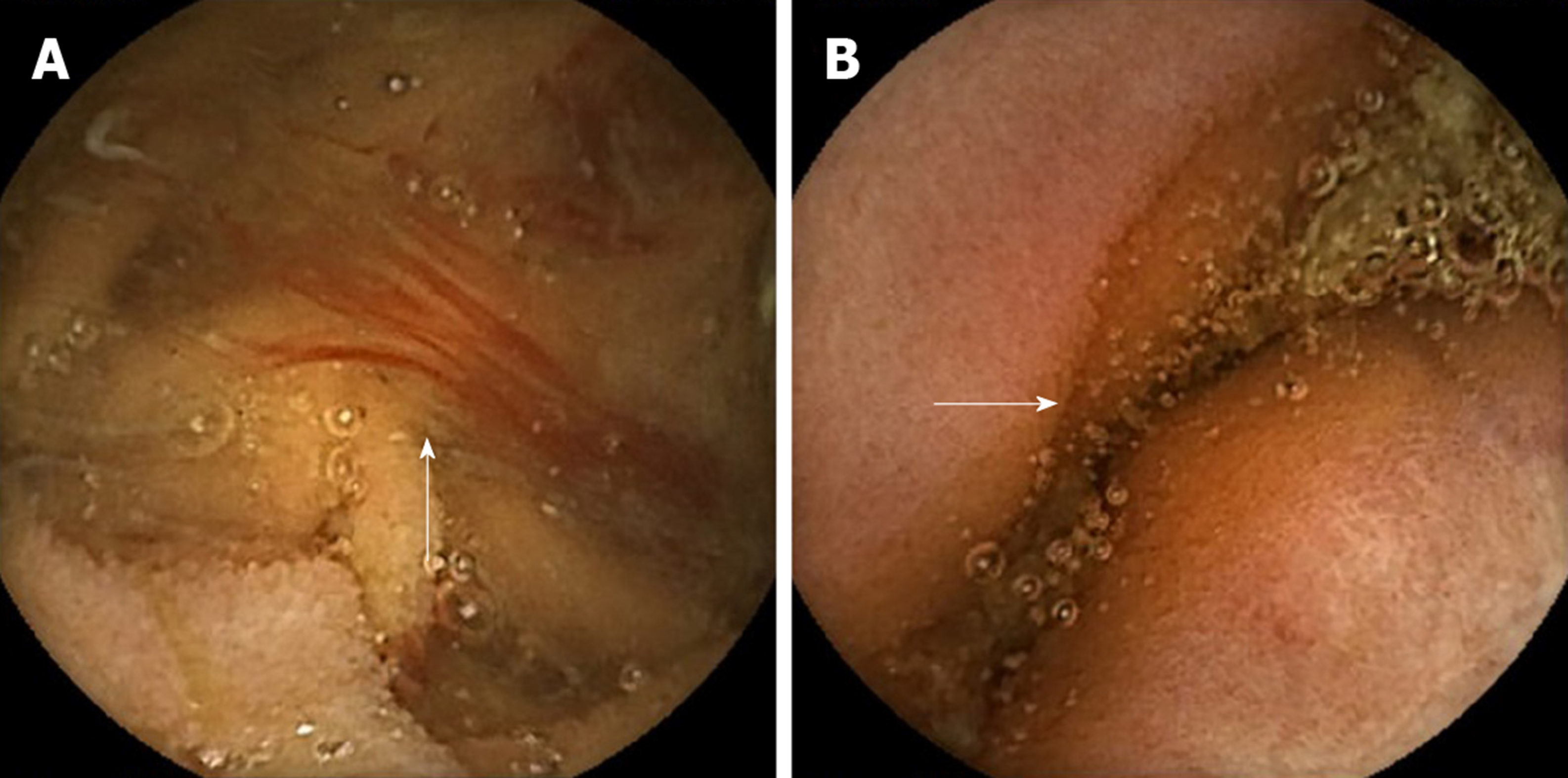
Figure 1 Videocapsule endoscopy findings obtained from our patient.
A: Fresh blood in the jejunum; B: Protruding jejunal lesion.
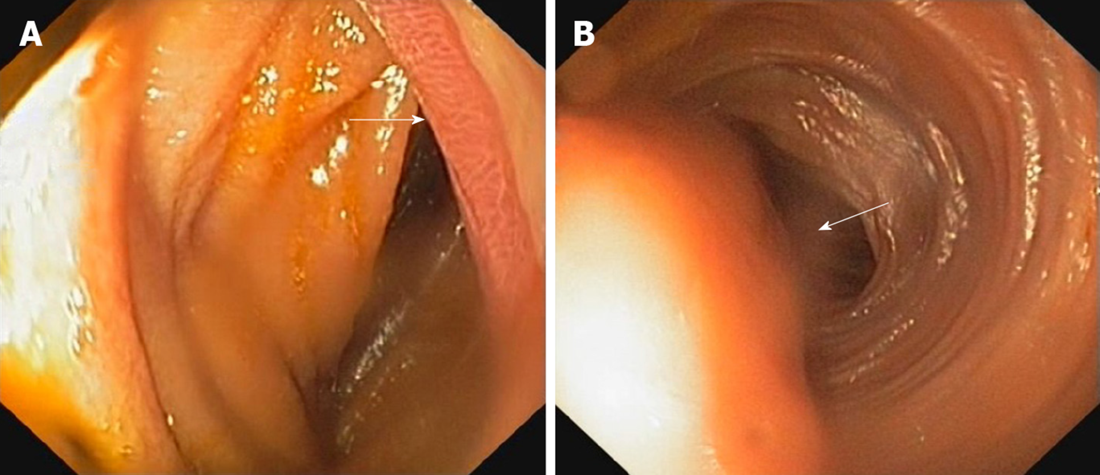
Figure 2 Images acquired through enteroscopy performed in our patient.
A: Ulcerated tumoral mass; B: Tumoral mass with partial bowel obstruction.
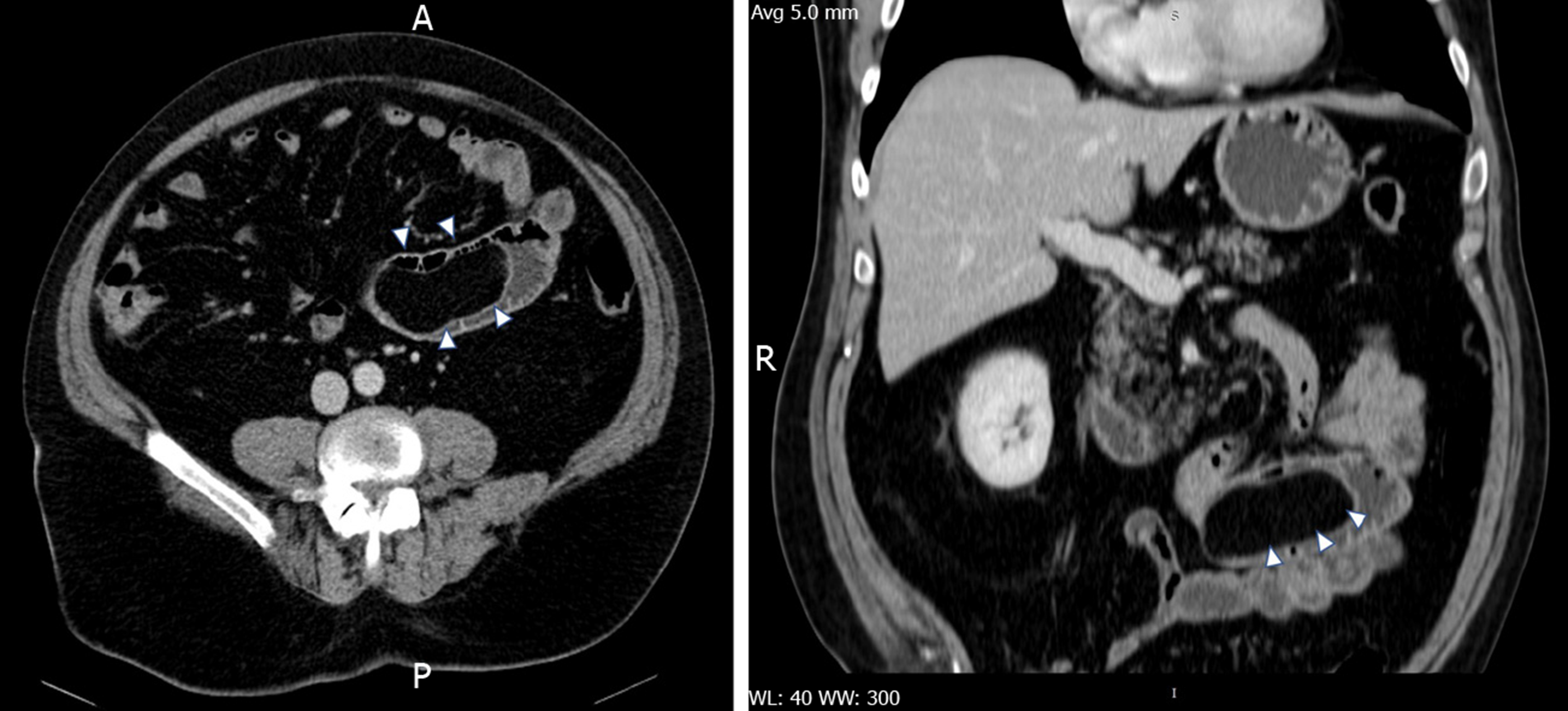
Figure 3 Contrast–enhanced abdominal computed tomography scan.
Both axial (left) and coronal (right) reformatted images show a large elongated structure inside of intestinal lumen (arrowheads) with homogeneous fat density and smooth, well defined contour.
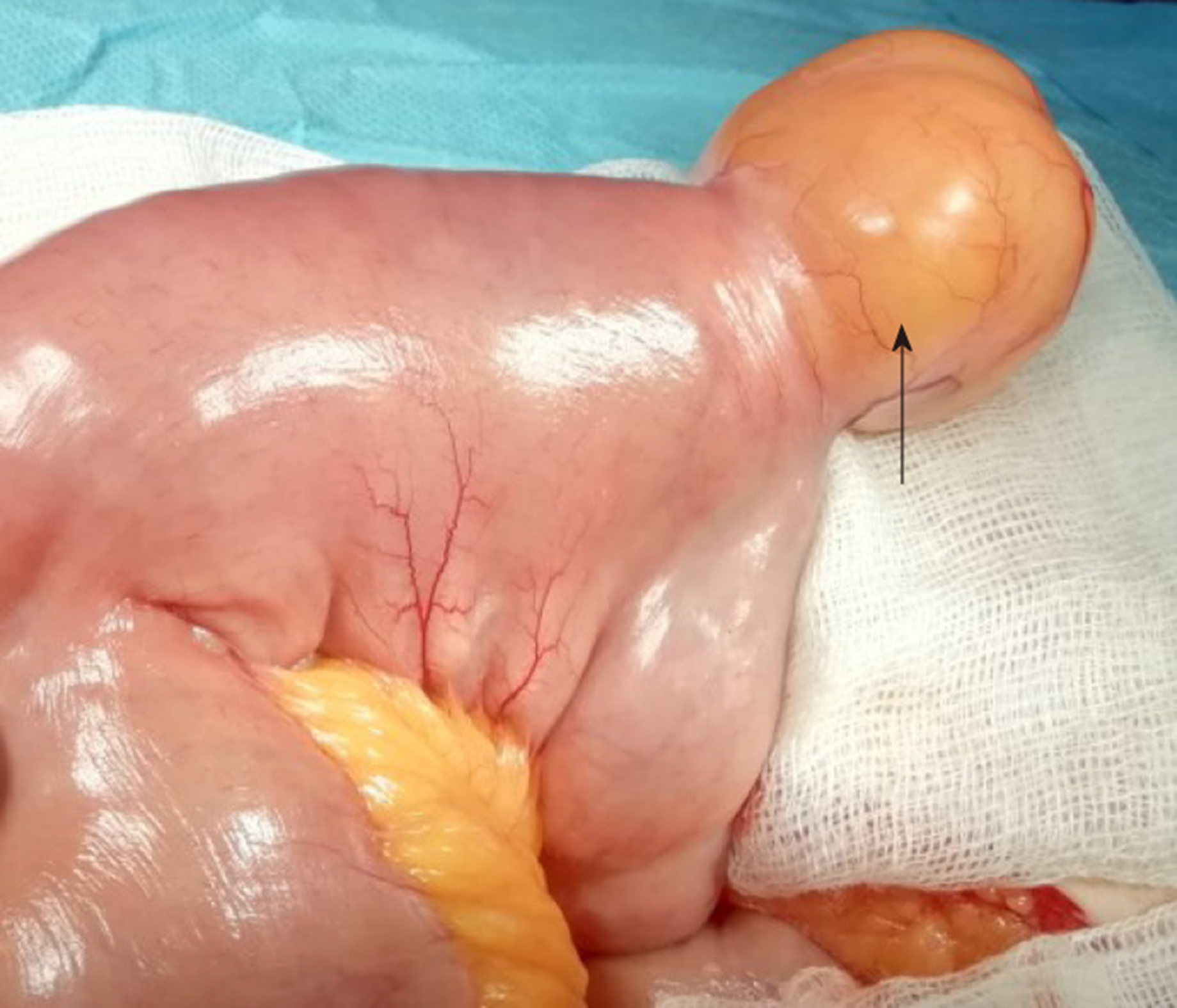
Figure 4 Macroscopic appearance of the jejunal lipoma (arrow).
Figure 5 Jejunal submucosal lipoma with ulcerated area of the mucosa.
A: Full section (Hematoxylin-eosin staining, × 40); B: Detail (Hematoxylin-eosin staining, × 200).
- Citation: Cuciureanu T, Huiban L, Chiriac S, Singeap AM, Danciu M, Mihai F, Stanciu C, Trifan A, Vlad N. Ulcerated intussuscepted jejunal lipoma-uncommon cause of obscure gastrointestinal bleeding: A case report. World J Clin Cases 2019; 7(22): 3765-3771
- URL: https://www.wjgnet.com/2307-8960/full/v7/i22/3765.htm
- DOI: https://dx.doi.org/10.12998/wjcc.v7.i22.3765


