Copyright
©The Author(s) 2019.
World J Clin Cases. Nov 6, 2019; 7(21): 3662-3670
Published online Nov 6, 2019. doi: 10.12998/wjcc.v7.i21.3662
Published online Nov 6, 2019. doi: 10.12998/wjcc.v7.i21.3662
Figure 1 Computed tomography of the brain showed multiple calcifications in the dentate nucleus and basal ganglia of bilateral cerebellum hemispheres.
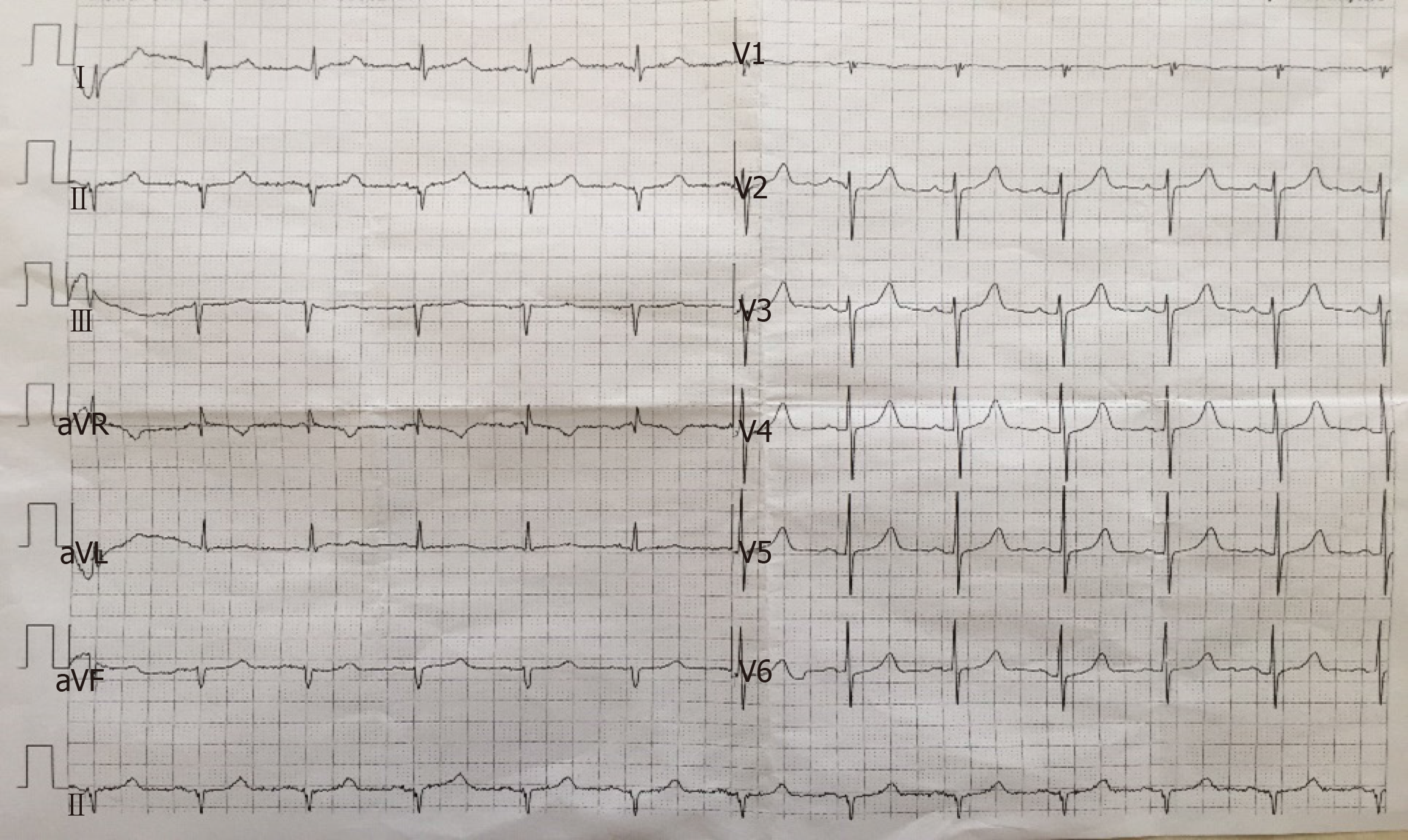
Figure 2 Electrocardiogram showing: (1) Sinus rhythm; (2) Electrocardiogram axis shifting -61° to the left; and (3) QS type in the II, III, and avF leads.
- Citation: Zhou YY, Yang Y, Qiu HM. Hypoparathyroidism with Fahr’s syndrome: A case report and review of the literature. World J Clin Cases 2019; 7(21): 3662-3670
- URL: https://www.wjgnet.com/2307-8960/full/v7/i21/3662.htm
- DOI: https://dx.doi.org/10.12998/wjcc.v7.i21.3662


