Copyright
©The Author(s) 2019.
World J Clin Cases. Oct 26, 2019; 7(20): 3347-3352
Published online Oct 26, 2019. doi: 10.12998/wjcc.v7.i20.3347
Published online Oct 26, 2019. doi: 10.12998/wjcc.v7.i20.3347
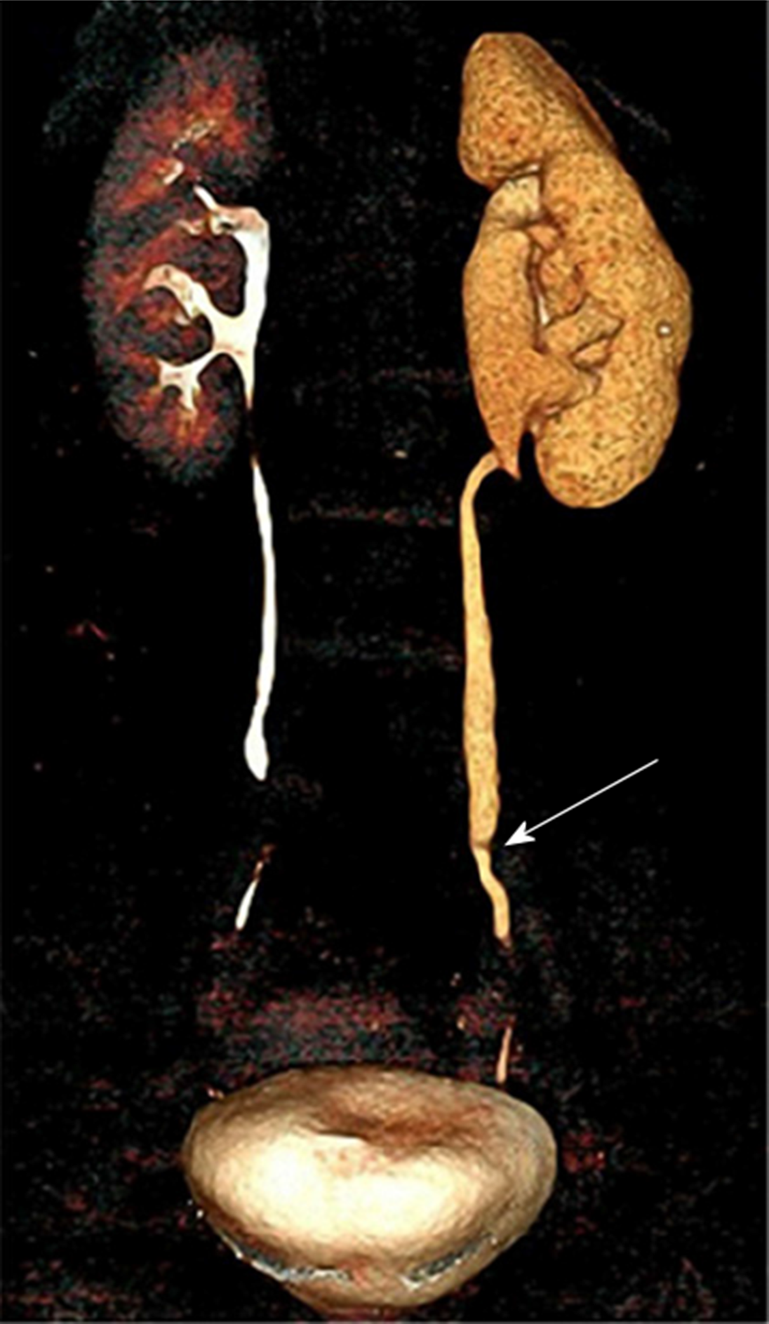
Figure 1 Computed tomography urography scan showed a severe obstruction in the lower segment of the left ureter along with left hydronephrosis.
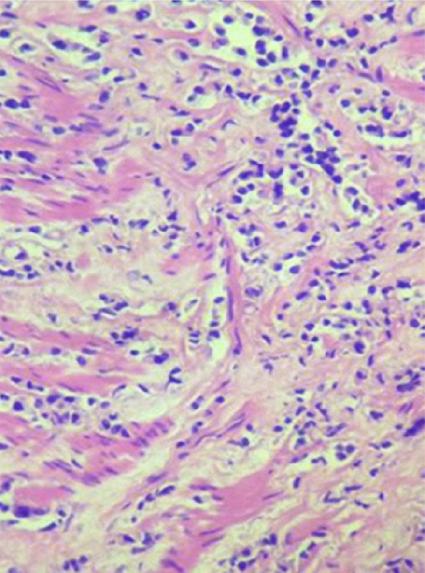
Figure 2 Hematoxylin and eosin-stained histological ureter section showing infiltration of poorly differentiated tumor cells that displayed an alveolar growth pattern (magnification, 100×).
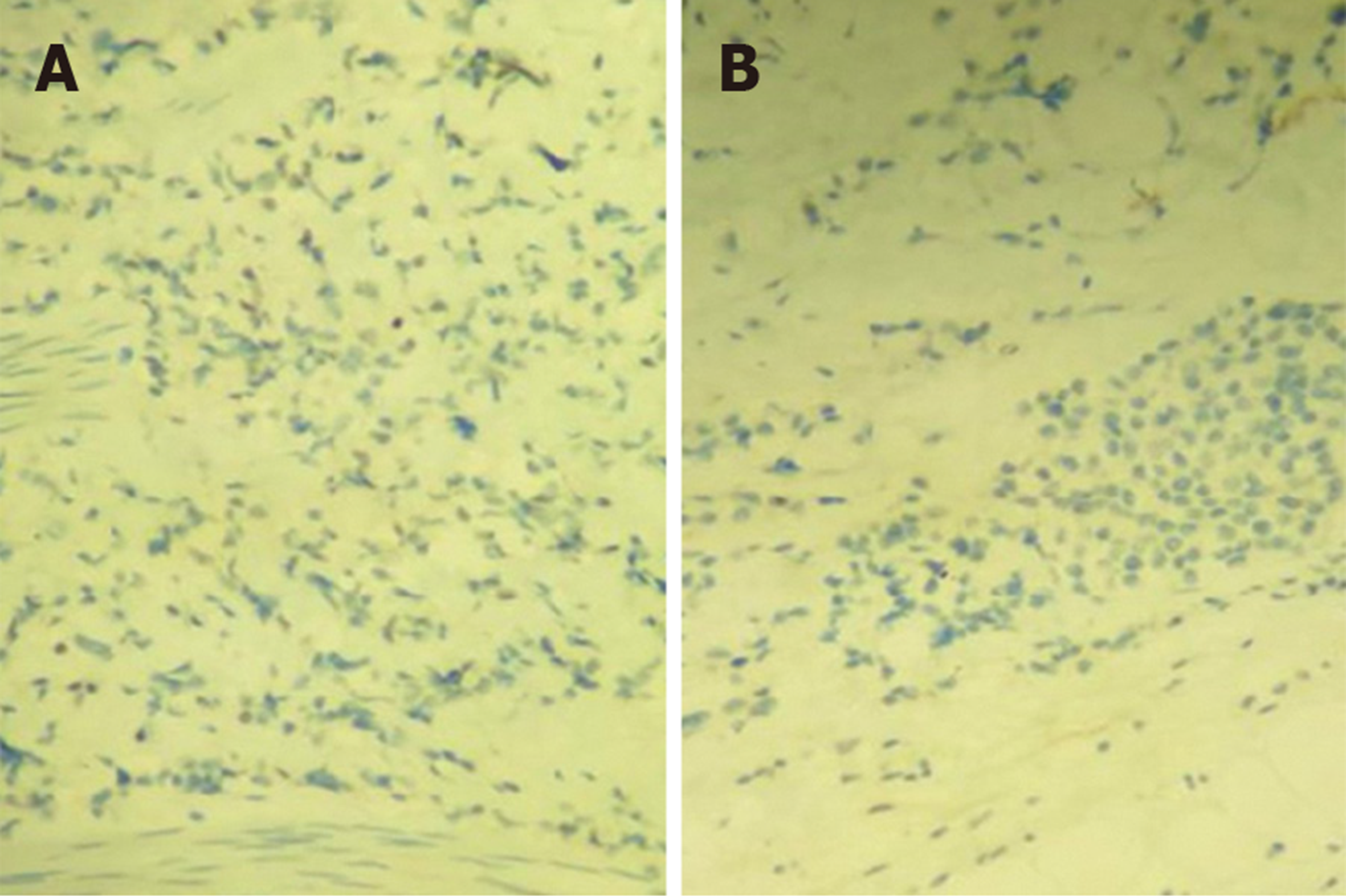
Figure 3 Immunoreactivity in tumor cells.
A: Weak nuclear immunoreactivity for estrogen receptor in tumor cells (magnification, 100×); B: Negative immunohistochemical staining for progestin receptor in tumor cells (magnification, 100×).
- Citation: Zhou ZH, Sun LJ, Zhang GM. Ureter - an unusual site of breast cancer metastasis: A case report. World J Clin Cases 2019; 7(20): 3347-3352
- URL: https://www.wjgnet.com/2307-8960/full/v7/i20/3347.htm
- DOI: https://dx.doi.org/10.12998/wjcc.v7.i20.3347


