Copyright
©The Author(s) 2019.
World J Clin Cases. Oct 26, 2019; 7(20): 3335-3340
Published online Oct 26, 2019. doi: 10.12998/wjcc.v7.i20.3335
Published online Oct 26, 2019. doi: 10.12998/wjcc.v7.i20.3335
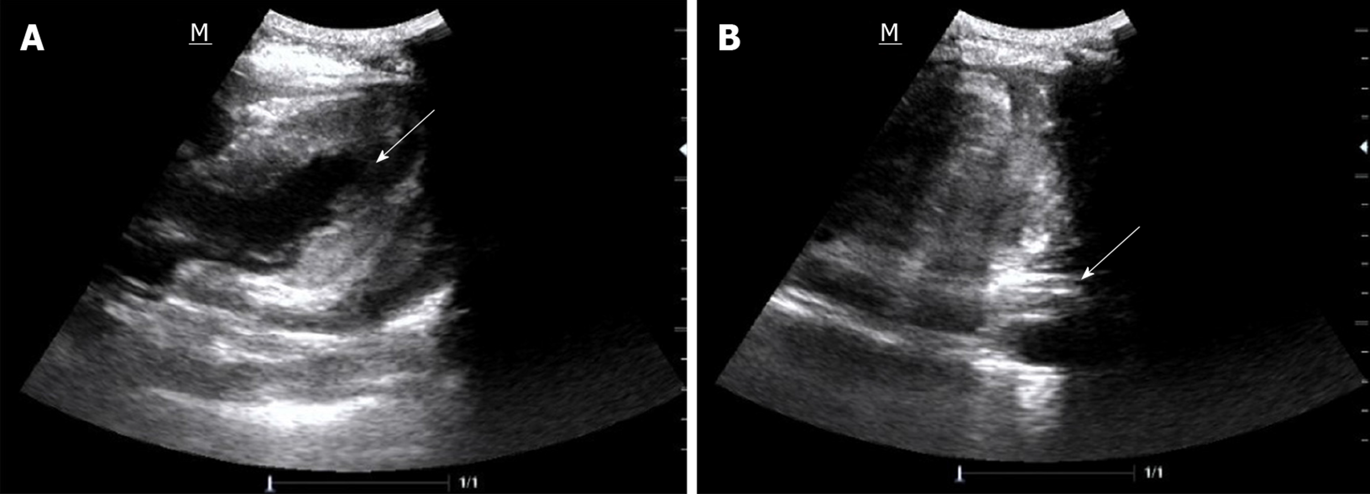
Figure 1 Bedside cardiac ultrasound images.
A: Pericardial effusion (arrow); B: A large number of pericardial blood clots (arrow).
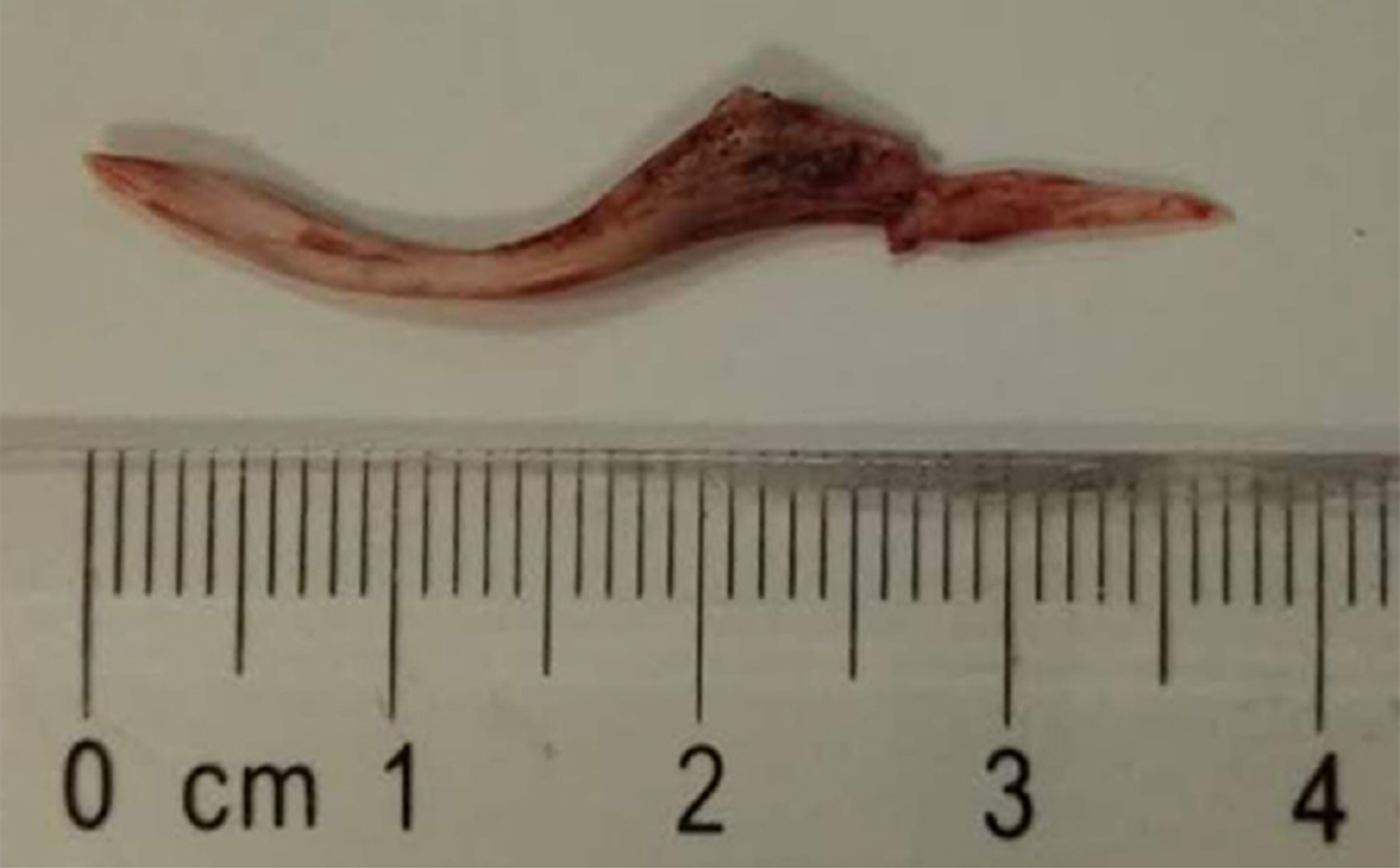
Figure 2 The fish bone 3.
7 cm in size.
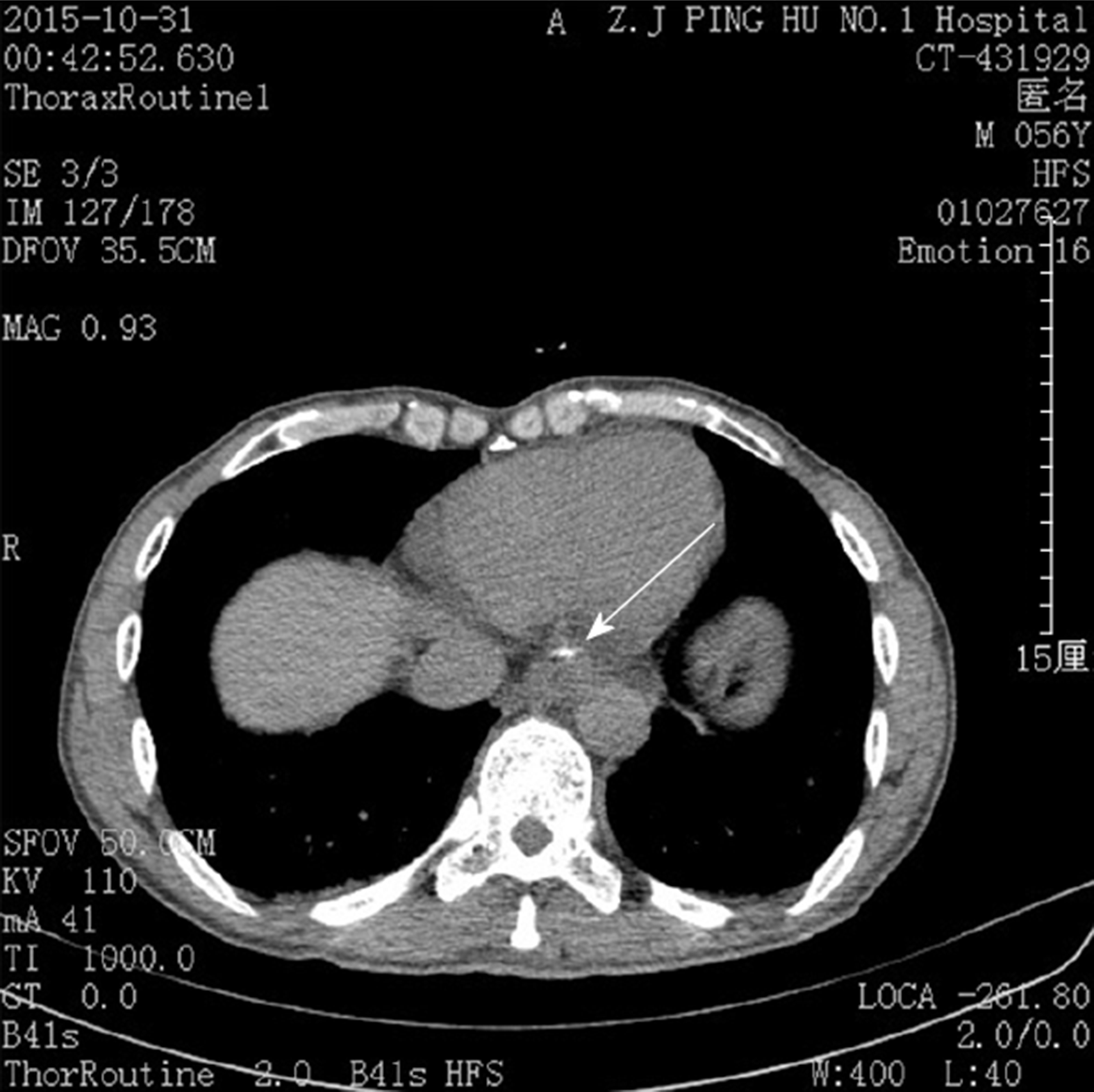
Figure 3 Chest computed tomography image.
A strip-shaped high-density shadow was visible at the bottom of the esophagus (arrow).
- Citation: Wang QQ, Hu Y, Zhu LF, Zhu WJ, Shen P. Fish bone-induced myocardial injury leading to a misdiagnosis of acute myocardial infarction: A case report. World J Clin Cases 2019; 7(20): 3335-3340
- URL: https://www.wjgnet.com/2307-8960/full/v7/i20/3335.htm
- DOI: https://dx.doi.org/10.12998/wjcc.v7.i20.3335


