Copyright
©The Author(s) 2019.
World J Clin Cases. Oct 26, 2019; 7(20): 3316-3321
Published online Oct 26, 2019. doi: 10.12998/wjcc.v7.i20.3316
Published online Oct 26, 2019. doi: 10.12998/wjcc.v7.i20.3316
Figure 1 Imaging findings of the head of the pancreas.
A: A non-uniformly enhanced soft tissue mass in the head of the pancreas on enhanced computed tomography (CT); B: The mass of the pancreatic head is hypointense on T1-weighted imaging (T1WI); C: The pancreatic head mass on diffusion-weighted imaging (DWI) is hyperintense; D: Contrast-enhanced magnetic resonance imaging (MRI) showed spoke wheel-like enhancement at the edge of the pancreatic head mass.
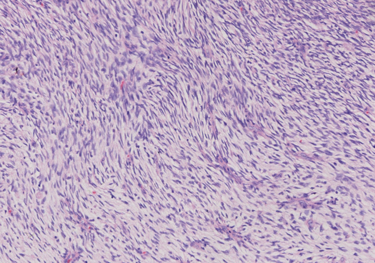
Figure 2 Postoperative pathologic photomicrograph showing a mesenchymal neoplasm in which the tumor and matrix cells were interwoven (HE staining, 200×).
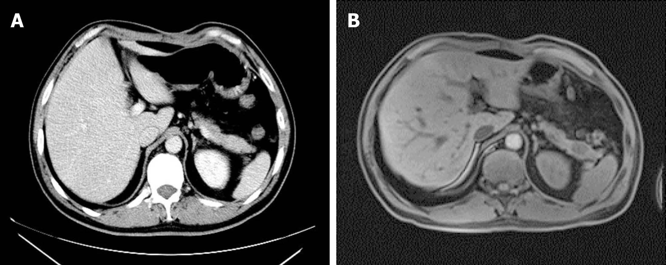
Figure 3 Imaging findings of the pancreatic tail.
A: Contrast-enhanced computed tomography showed no abnormal mass in the pancreatic tail; B: Enhanced magnetic resonance imaging clearly showed the mass at the tail of the pancreas.
- Citation: Cai HJ, Fang JH, Cao N, Wang W, Kong FL, Sun XX, Huang B. Dermatofibrosarcoma metastases to the pancreas: A case report. World J Clin Cases 2019; 7(20): 3316-3321
- URL: https://www.wjgnet.com/2307-8960/full/v7/i20/3316.htm
- DOI: https://dx.doi.org/10.12998/wjcc.v7.i20.3316


