Copyright
©The Author(s) 2019.
World J Clin Cases. Oct 26, 2019; 7(20): 3296-3302
Published online Oct 26, 2019. doi: 10.12998/wjcc.v7.i20.3296
Published online Oct 26, 2019. doi: 10.12998/wjcc.v7.i20.3296
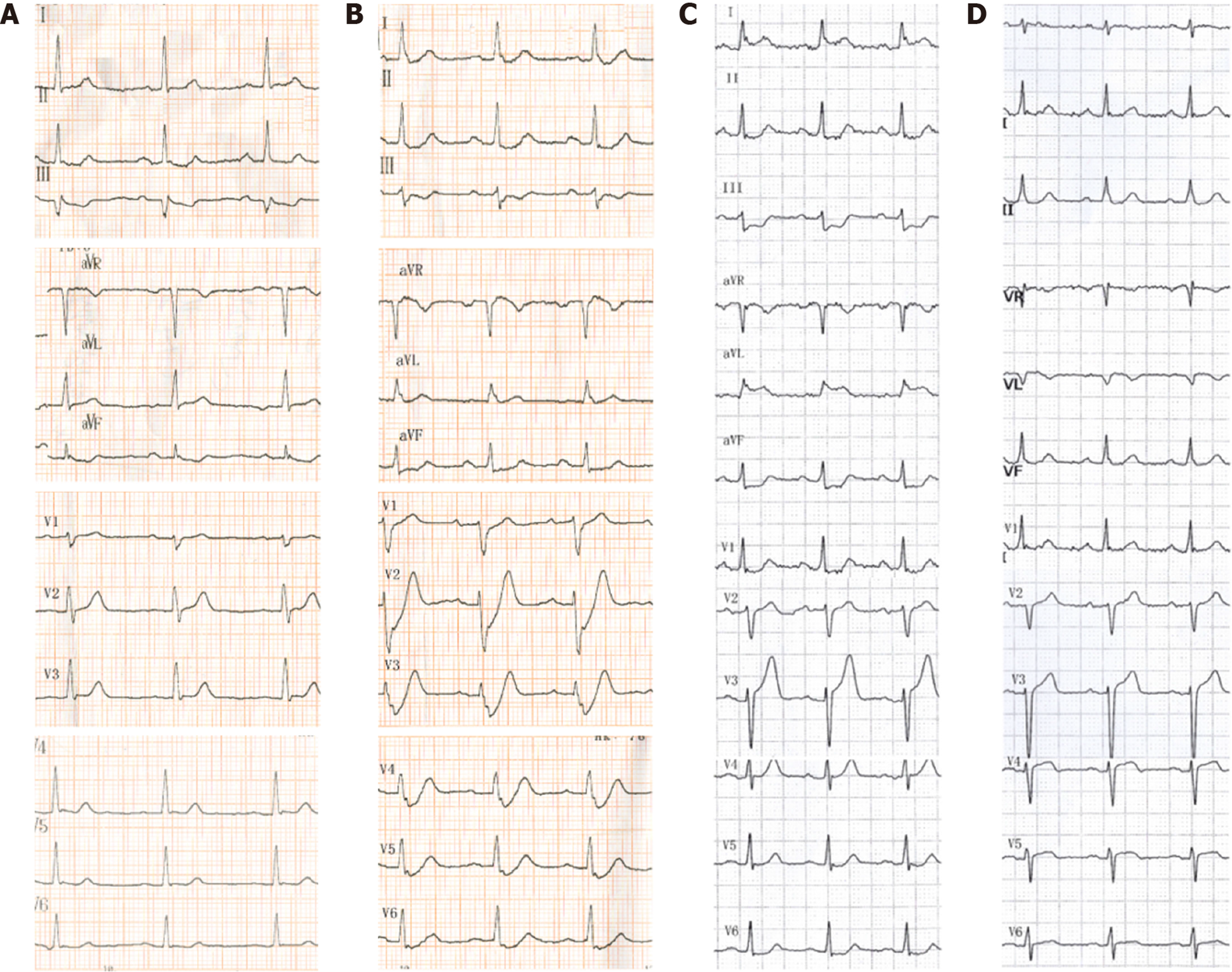
Figure 1 Electrocardiography examinations (Case 1).
A: Electrocardiogram performed 57 min after onset of pain; B: Electrocardiogram at the 100th min with persistent chest pain; C: Electrocardiogram at the 172th min with persistent chest pain; D: Electrocardiogram after the percutaneous coronary intervention.
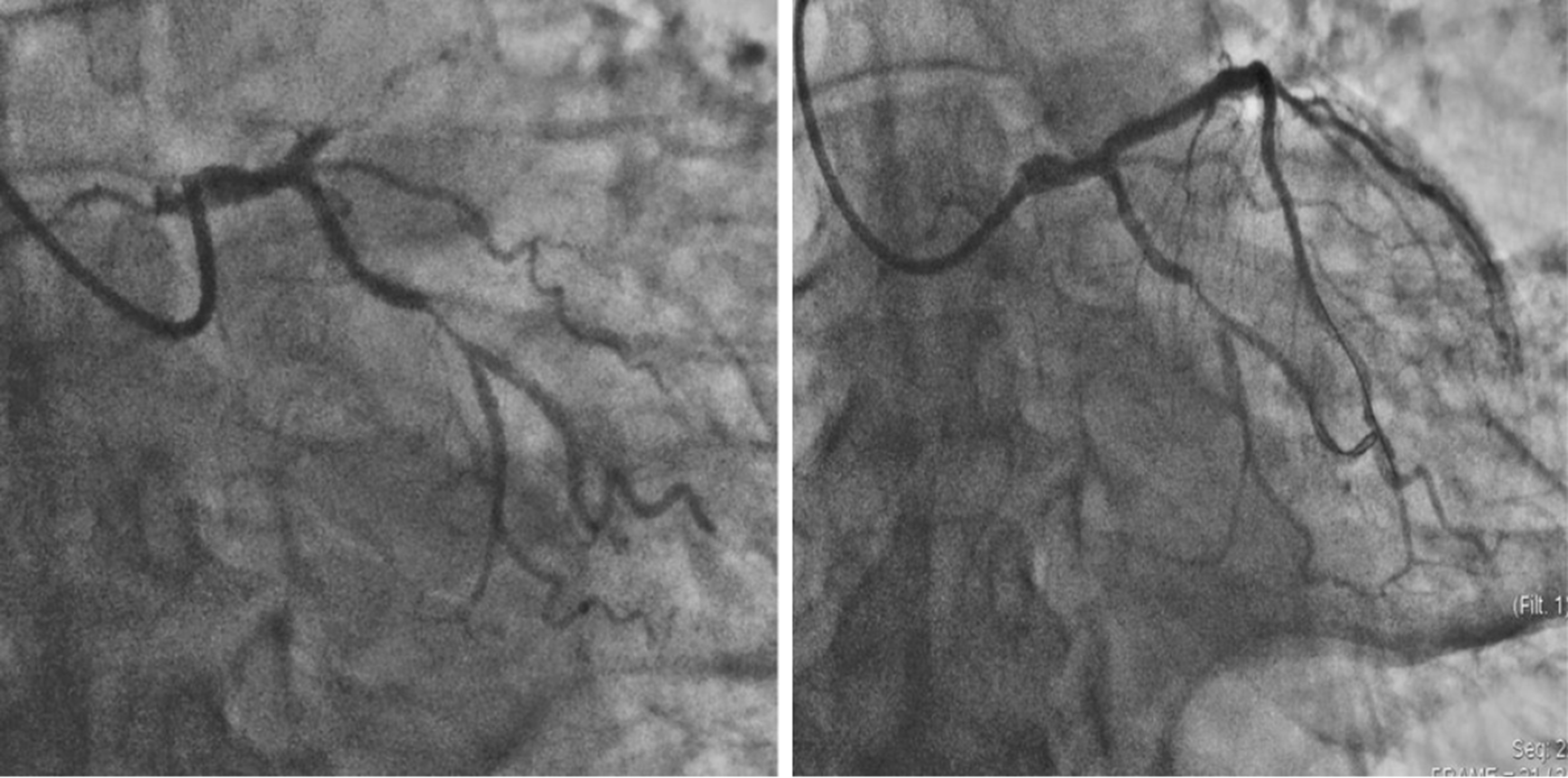
Figure 2 Coronary angiogram showing complete occlusion of the proximal left anterior descending coronary and a 90% stenosis of the middle of the left circumflex arteries (left) and that after drug-eluting stent placement (right).
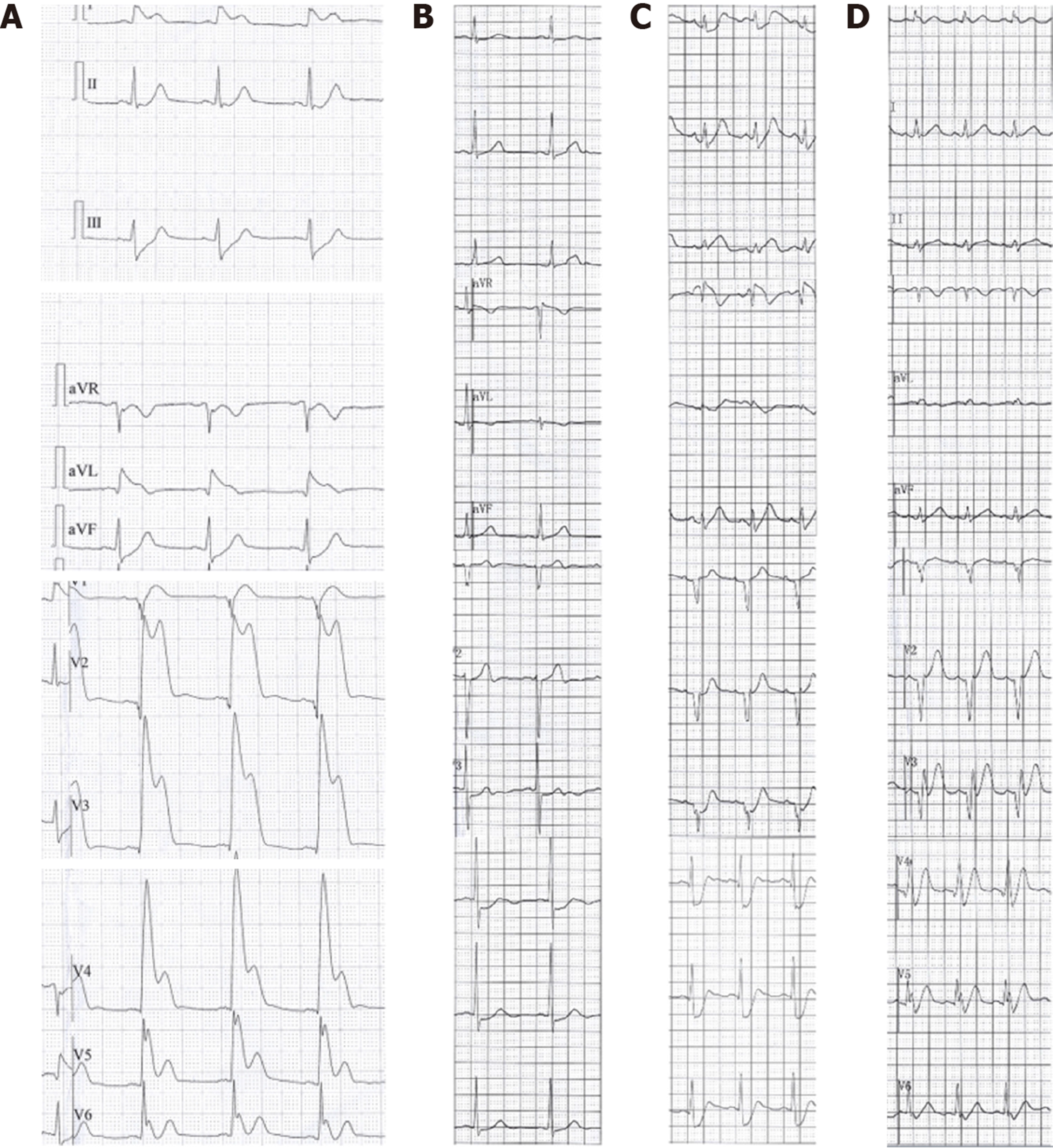
Figure 3 Electrocardiography examinations (Case 2).
A: Electrocardiogram at the 116th min after onset of chest pain; B: Electrocardiogram at the 155th min when symptoms alleviated; C: Electrocardiogram at the 236th min when chest pain recurred; D: Electrocardiogram recorded prior to emergency coronary angiography (at the 255th min after onset of pain).
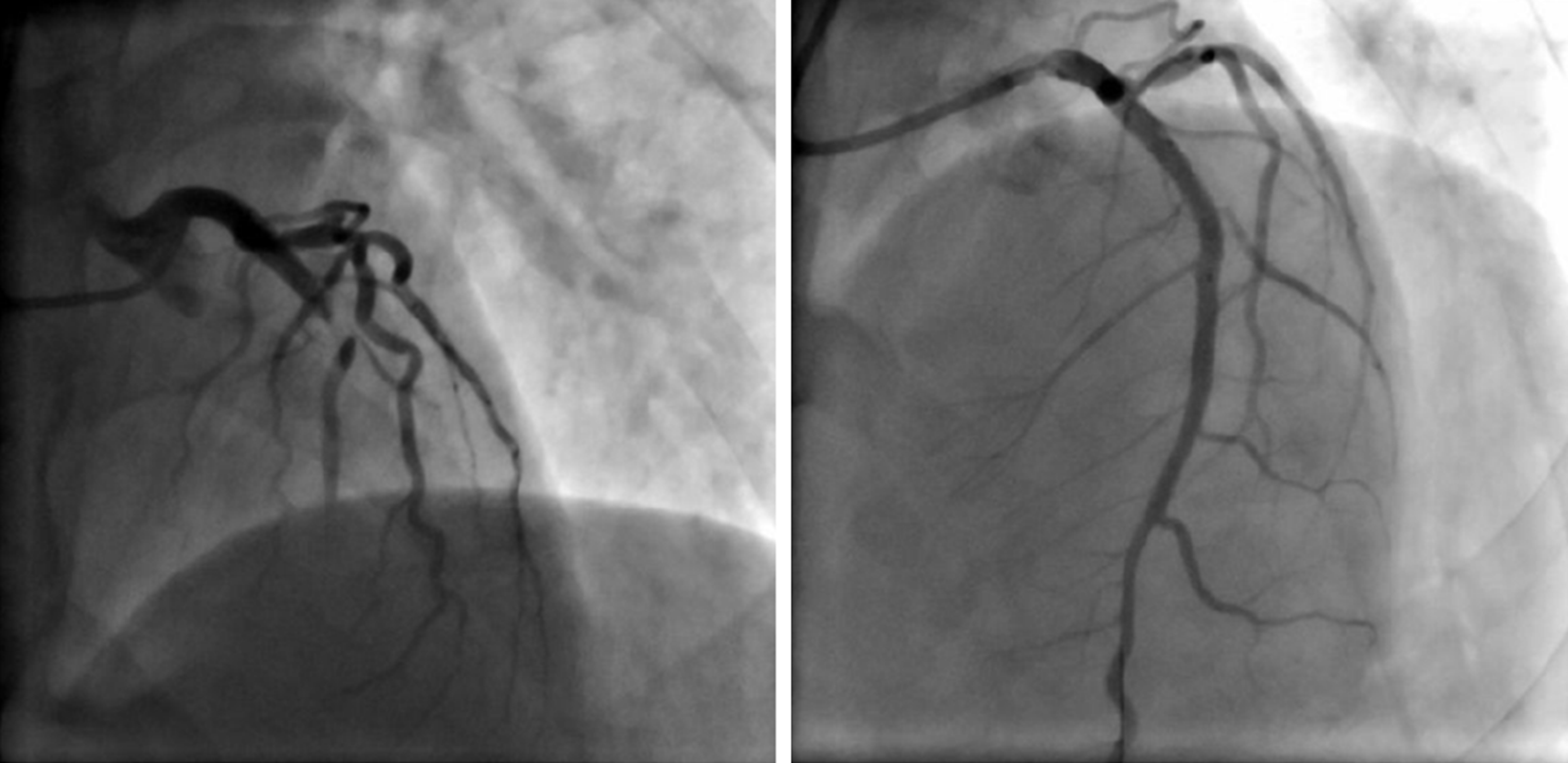
Figure 4 Coronary angiogram showing a 99% stenosis of the middle left anterior descending artery (left) and that after drug-eluting stent placement (right).
- Citation: Lin YY, Wen YD, Wu GL, Xu XD. De Winter syndrome and ST-segment elevation myocardial infarction can evolve into one another: Report of two cases. World J Clin Cases 2019; 7(20): 3296-3302
- URL: https://www.wjgnet.com/2307-8960/full/v7/i20/3296.htm
- DOI: https://dx.doi.org/10.12998/wjcc.v7.i20.3296


