Copyright
©The Author(s) 2019.
World J Clin Cases. Oct 26, 2019; 7(20): 3282-3288
Published online Oct 26, 2019. doi: 10.12998/wjcc.v7.i20.3282
Published online Oct 26, 2019. doi: 10.12998/wjcc.v7.i20.3282
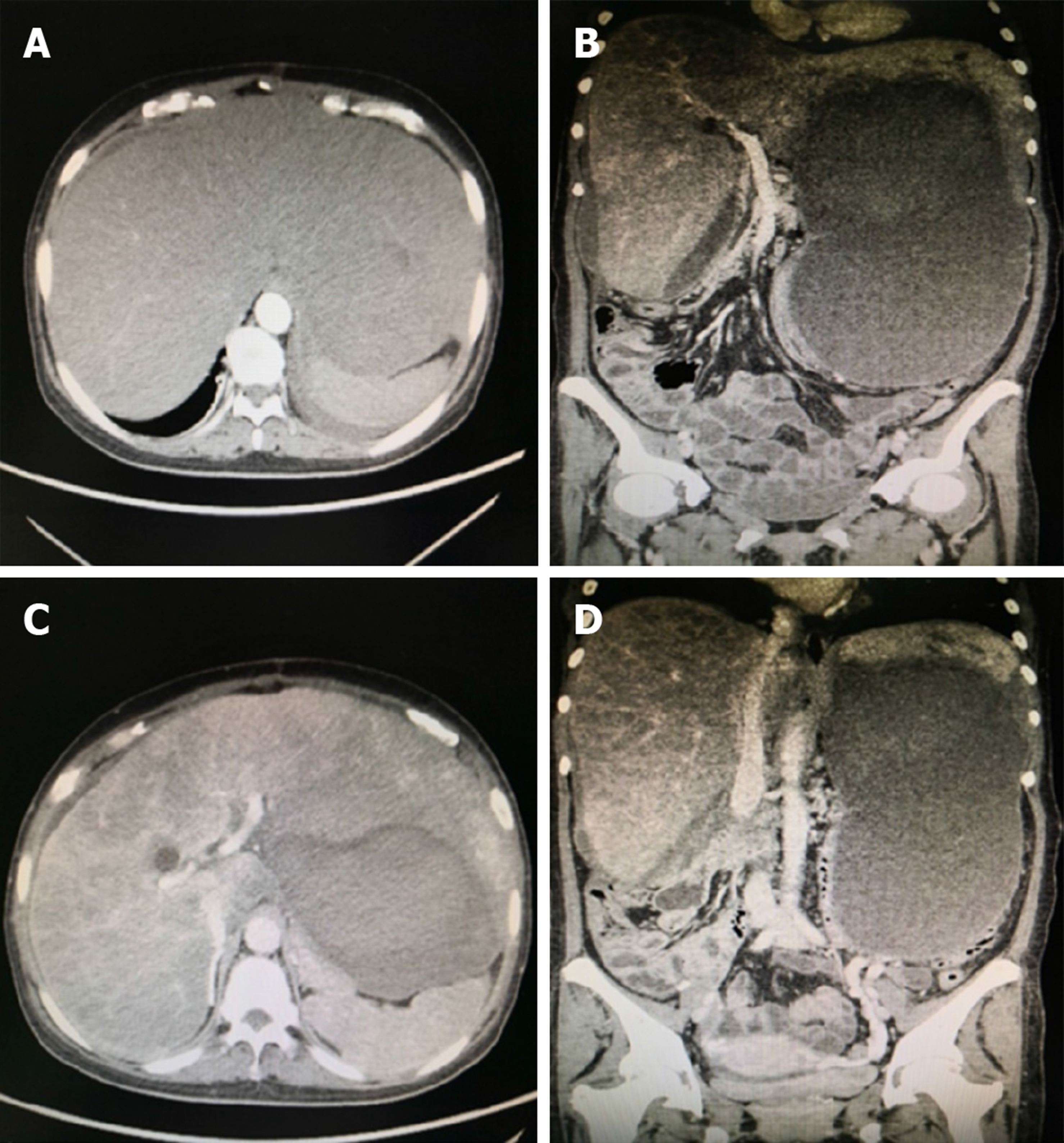
Figure 1 Abdominal computed tomography.
A: Hepatomegaly; B: Portal vein was not thick, and no filling defect was observed; C: Three hepatic veins were not seen; D: Stenosis of the inferior vena cava of the hepatic segment and a huge hematoma in the liver.
Figure 2 The contrast agent entered the inferior vena cava through the stents and the systemic circulation.
A: Portal vein; B, C: The shunt between the portal vein and the inferior vena cava.
Figure 3 The inferior vena cava of the hepatic segment was still narrowed but to a lesser extent.
The three hepatic veins could not be seen.
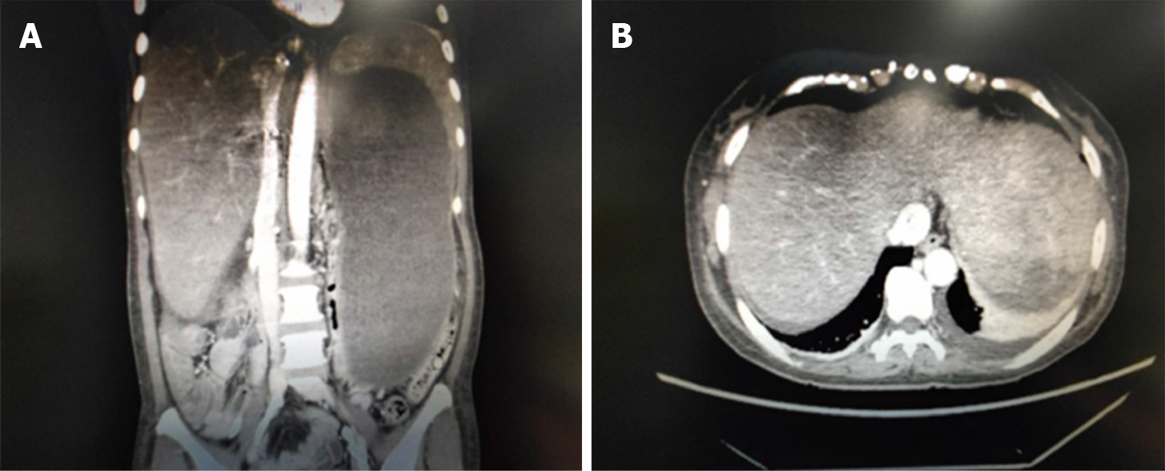
Figure 4 The inferior vena cava of the hepatic segment was obviously expanded.
The three hepatic veins were still not seen.
- Citation: Li TT, Wu YF, Liu FQ, He FL. Hepatic amyloidosis leading to hepatic venular occlusive disease and Budd-Chiari syndrome: A case report. World J Clin Cases 2019; 7(20): 3282-3288
- URL: https://www.wjgnet.com/2307-8960/full/v7/i20/3282.htm
- DOI: https://dx.doi.org/10.12998/wjcc.v7.i20.3282


