Copyright
©The Author(s) 2019.
World J Clin Cases. Oct 26, 2019; 7(20): 3271-3275
Published online Oct 26, 2019. doi: 10.12998/wjcc.v7.i20.3271
Published online Oct 26, 2019. doi: 10.12998/wjcc.v7.i20.3271
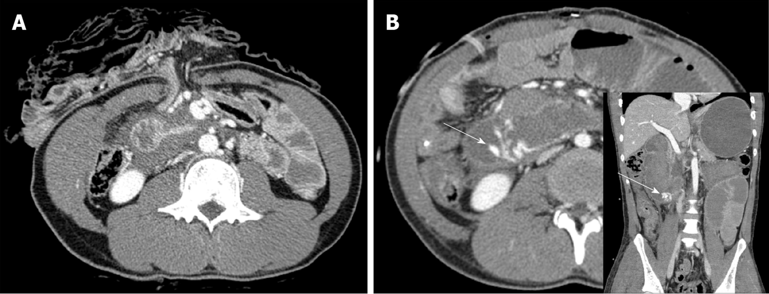
Figure 1 Abdominal computed tomogrpahy.
A: Preoperative abdominal computed tomogrpahy (CT) shows paraduodenal fluid collection and transverse colon and small bowel herniation from the abdominal stab wound; B: Postoperative abdominopelvic CT shows extravasation of the contrast medium in the third portion of the duoenum (arrow).
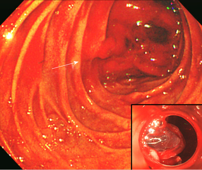
Figure 2 Endoscopy showed active bleeding with a perforation in the third portion of the duodenum.
The cap-fitted endoscopy enabled closer observation of the bleeding site.
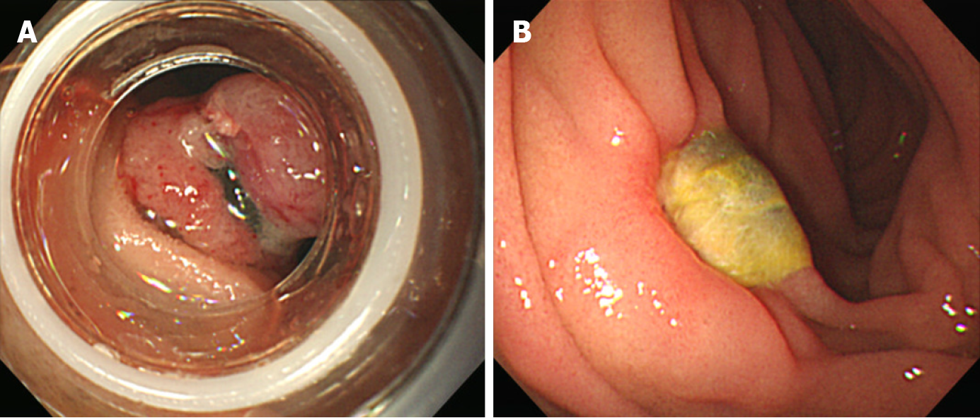
Figure 3 Endoscopy results.
A: Endoscopic band ligation was used to make a mushroom-like area of mucosa to obtain complete closure. Additional band ligation was performed to get a more stable anchor; B: Follow-up endoscopy 9 d later showed a healing ulcer at the previous site of bleeding.
- Citation: Kim DH, Choi H, Kim KB, Yun HY, Han JH. Endoluminal closure of an unrecognized penetrating stab wound of the duodenum with endoscopic band ligation: A case report. World J Clin Cases 2019; 7(20): 3271-3275
- URL: https://www.wjgnet.com/2307-8960/full/v7/i20/3271.htm
- DOI: https://dx.doi.org/10.12998/wjcc.v7.i20.3271


