Copyright
©The Author(s) 2019.
World J Clin Cases. Jan 26, 2019; 7(2): 191-202
Published online Jan 26, 2019. doi: 10.12998/wjcc.v7.i2.191
Published online Jan 26, 2019. doi: 10.12998/wjcc.v7.i2.191
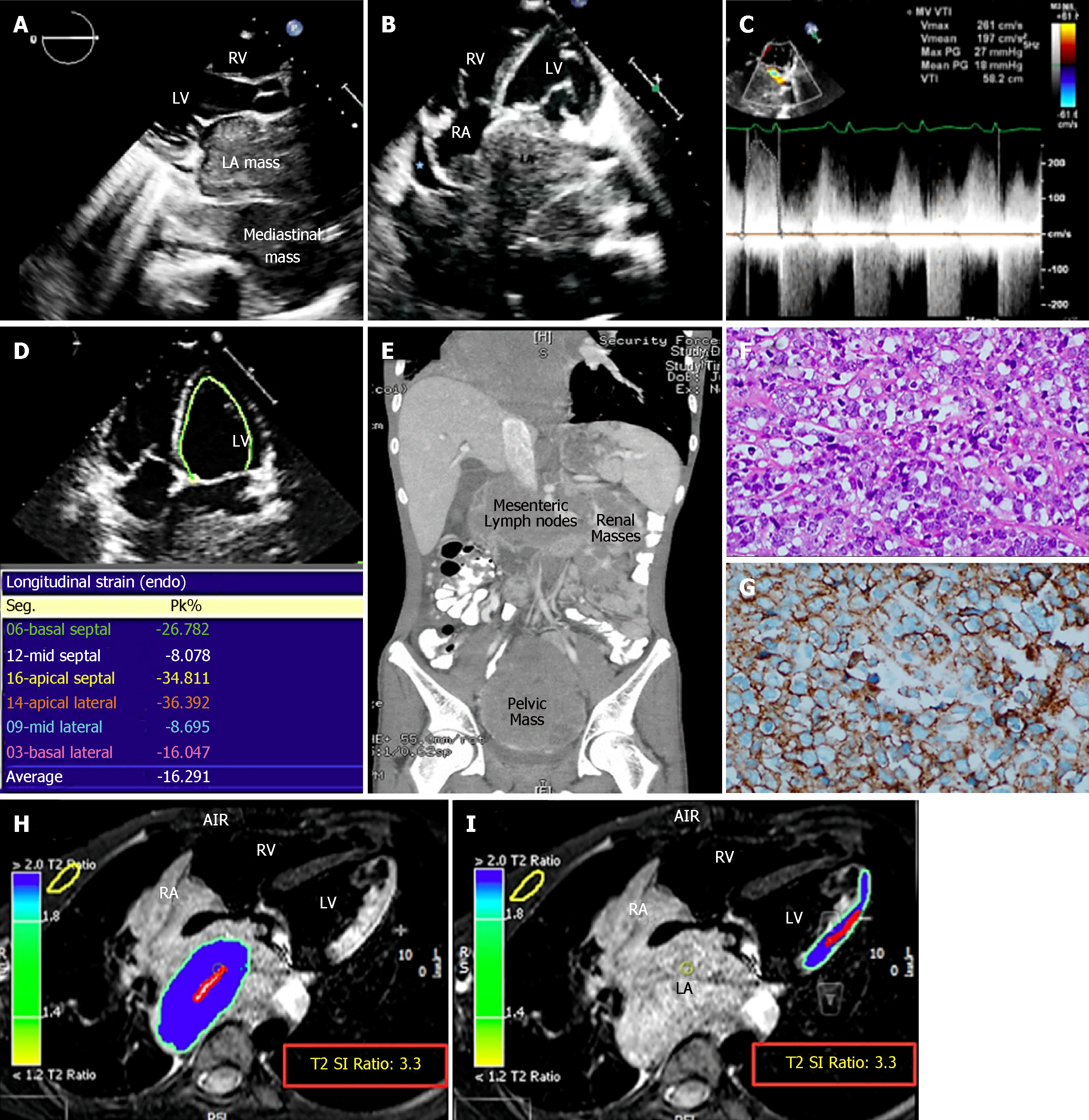
Figure 1 The patient’s initial imaging and histopathological findings.
A and B: Echocardiographic findings: (A) Parasternal long axis and (B) apical 4-chamber views demonstrating a very large LA mass extending from the mediastinum and involving the RA. Minimal pericardial effusion is noted (blue star); C: Continuous-wave Doppler showing functional mitral stenosis and regurgitation; D: Strain imaging of the LV in a 4-chamber view showing impaired lateral wall strain due to lymphoma infiltration; E: Contrast-enhanced thoracoabdominal computed tomography (CT) scan in a coronal view showing mesenteric, renal, and pelvic masses; F: Histopathology of the pelvic mass revealed atypical large lymphoid cells with prominent nucleoli and abundant cytoplasm; G: Immunohistochemistry was strongly positive for CD20; H and I: First CMRI images, STIR, axial views showing high signal intensity of the LA mass (H), and lateral LV wall (I), indicating high fluid content. The T2 signal intensity (SI) ratio of the LA mass and lateral LV wall are identical, indicating infiltration of the lateral LV wall by the mediastinal lymphoma. LA: Left atrium; RA: Right atrium; RV: Right ventricle; LV: Left ventricle; CMRI: Cardiac magnetic resonance imaging; STIR: T2-weighted short-tau inversion recovery images; SI: Signal intensity.
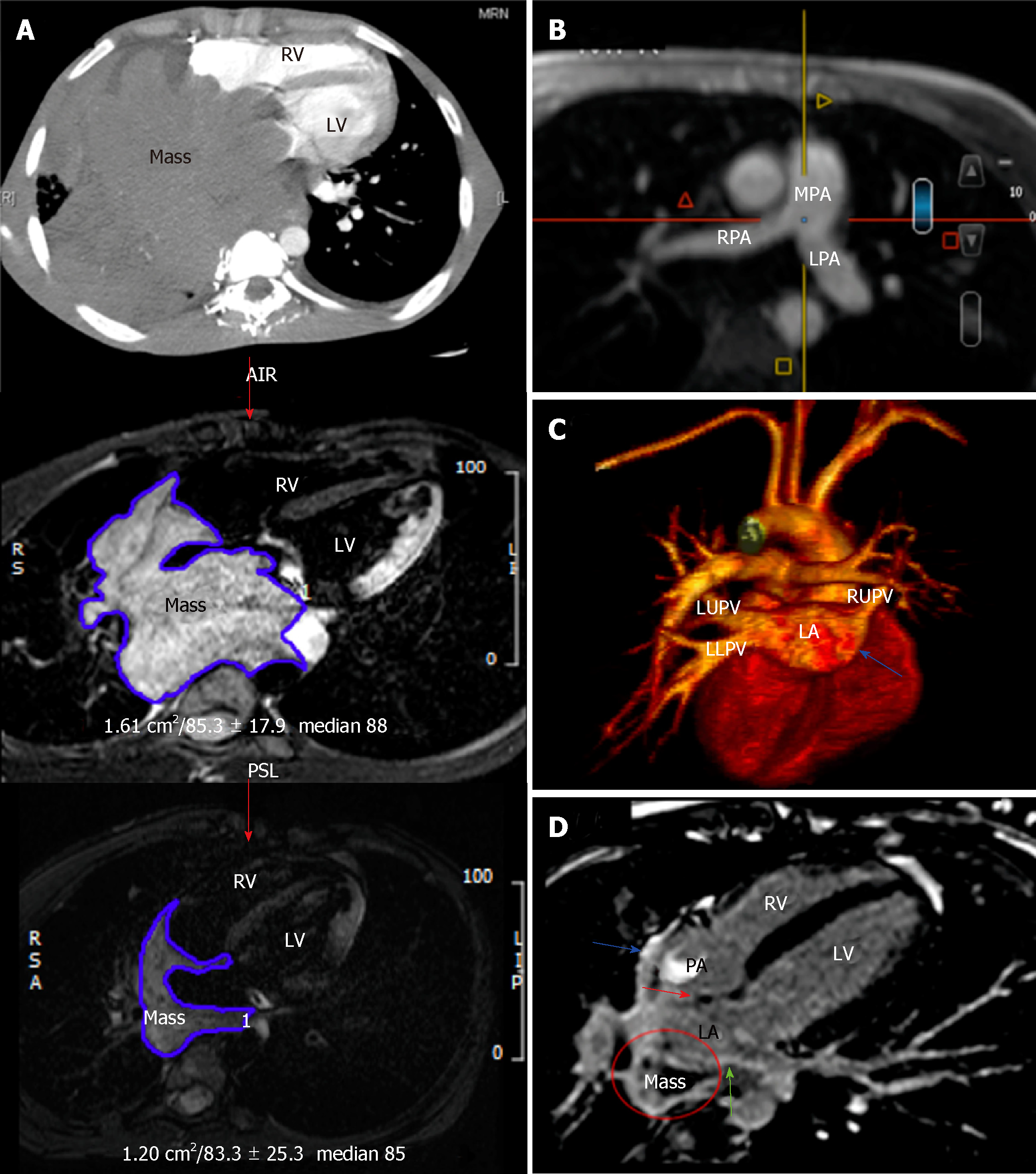
Figure 2 The patient’s imaging findings during the course of chemotherapy.
A: Follow-up computed tomography and CMRI at the heart level at consecutive time intervals showing gradual shrinkage of the mediastinal/cardiac mass size by 80% compared to baseline; B and C: Magnetic resonance angiography of the major cardiac vessels; B: Multiple-plane reformatted image showing the patency of the pulmonary arteries; C: In a 3-dimensional image, all pulmonary veins are now patent except the right lower pulmonary vein (blue arrow); D: Second CMRI-T1-weighted LGE image in a 4-chamber view revealing heterogeneous LGE of the mass (circle), suggesting tissue necrosis and fibrosis. The hyperintense signal of the interatrial septum (red arrow), left atrial (green arrow), and right atrial walls (blue arrows) suggest lymphoma resolution and healing by fibrosis. CMRI: Cardiac magnetic resonance imaging; RV: Right ventricle; LV: Left ventricle; RA: Right atrium; LA: Left atrium; LGE: Late gadolinium enhancement; MPA: Main pulmonary artery; RPA: Right pulmonary artery; LPA: Left pulmonary artery; LUPV: Left upper pulmonary vein; LLPV: Left lower pulmonary vein; RUPV: Right upper pulmonary vein.
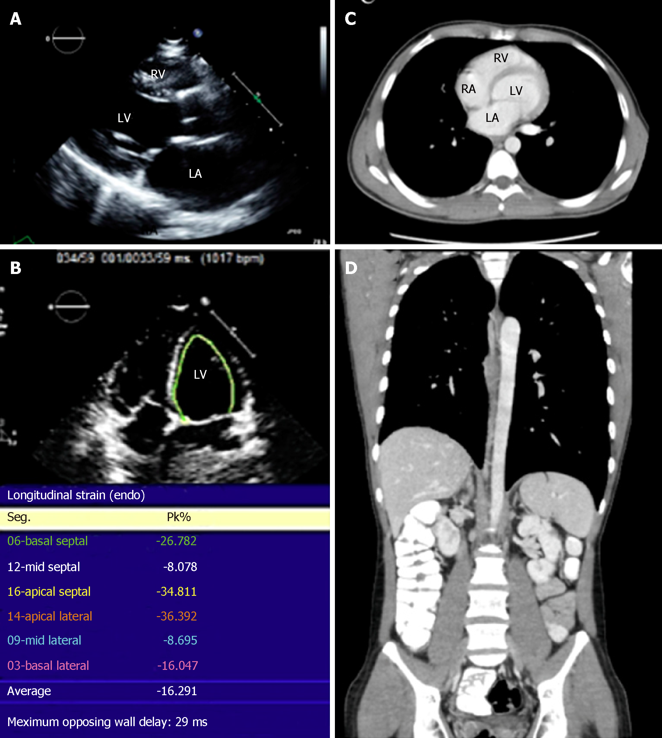
Figure 3 Recent imaging of the patient showing resolution of the lymphoma.
A: Normal parasternal long axis echocardiographic view; B: Strain imaging in a 4-chamber view showing improvement in lateral wall strain; C and D: Normal computed tomography images of the patient; C: Axial image of the chest; D: Coronal thoracoabdominal image. RV: Right ventricle; LV: Left ventricle; RA: Right atrium; LA: Left atrium.
- Citation: Al-Mehisen R, Al-Mohaissen M, Yousef H. Cardiac involvement in disseminated diffuse large B-cell lymphoma, successful management with chemotherapy dose reduction guided by cardiac imaging: A case report and review of literature. World J Clin Cases 2019; 7(2): 191-202
- URL: https://www.wjgnet.com/2307-8960/full/v7/i2/191.htm
- DOI: https://dx.doi.org/10.12998/wjcc.v7.i2.191


