Copyright
©The Author(s) 2019.
World J Clin Cases. Oct 6, 2019; 7(19): 3153-3159
Published online Oct 6, 2019. doi: 10.12998/wjcc.v7.i19.3153
Published online Oct 6, 2019. doi: 10.12998/wjcc.v7.i19.3153
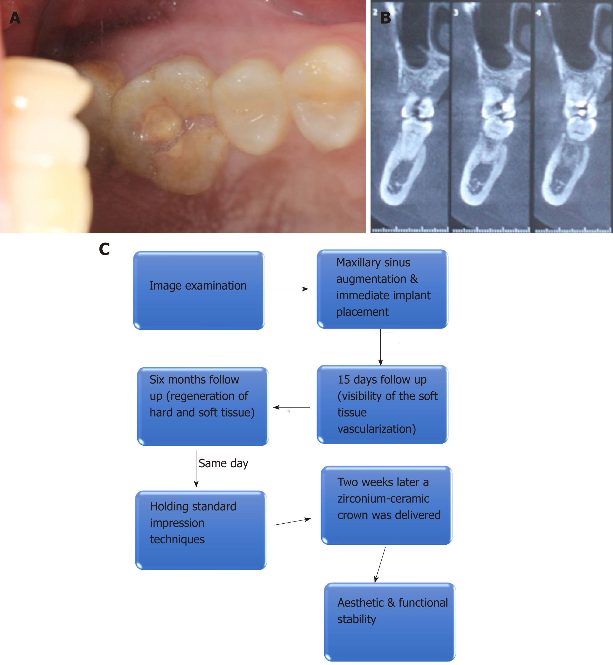
Figure 1 Preoperative intraoral condition and cone-beam computed tomography.
A: Tooth #26 presented with resin filling and vertical crown-root fracture; B: Cone-beam computed tomography revealed a fracture line from the occlusal surface to the end of the palatal root and the available bone height was 4 mm; C: Flow chart timeline of the treatment plan.
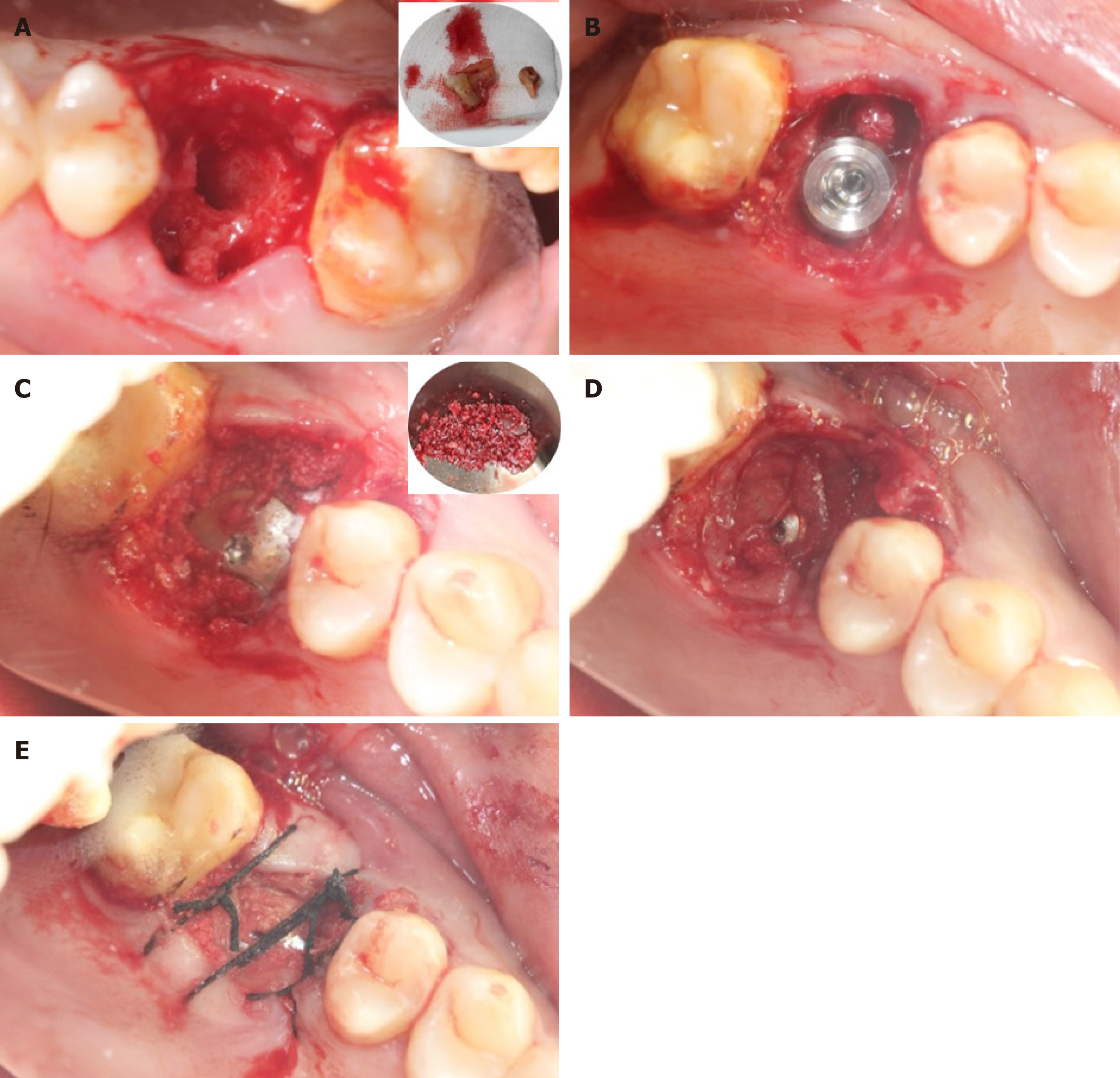
Figure 2 The surgical procedure.
A: The tooth #26 was extracted atraumatically; B: The implant was placed. The bone defect was visible around the implant; C: The bio-oss collagen with platelet rich fibrin (PRF) was placed into the space between the implant and the socket walls; D and E: The wound was covered with PRF membrane without tight suture to regenerate soft tissue.
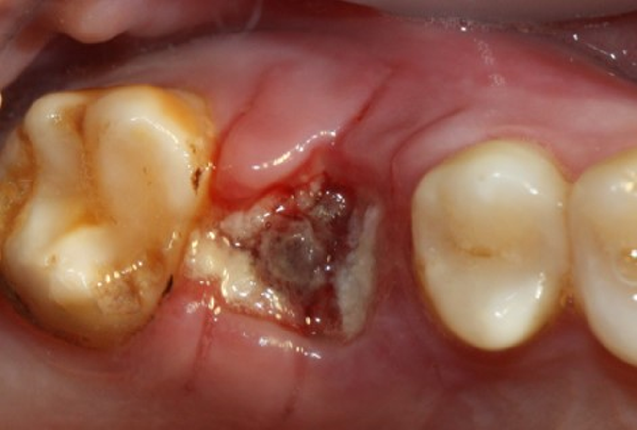
Figure 3 Intraoral condition at 15-d follow-up visit: The vascularization of soft tissue is visible.
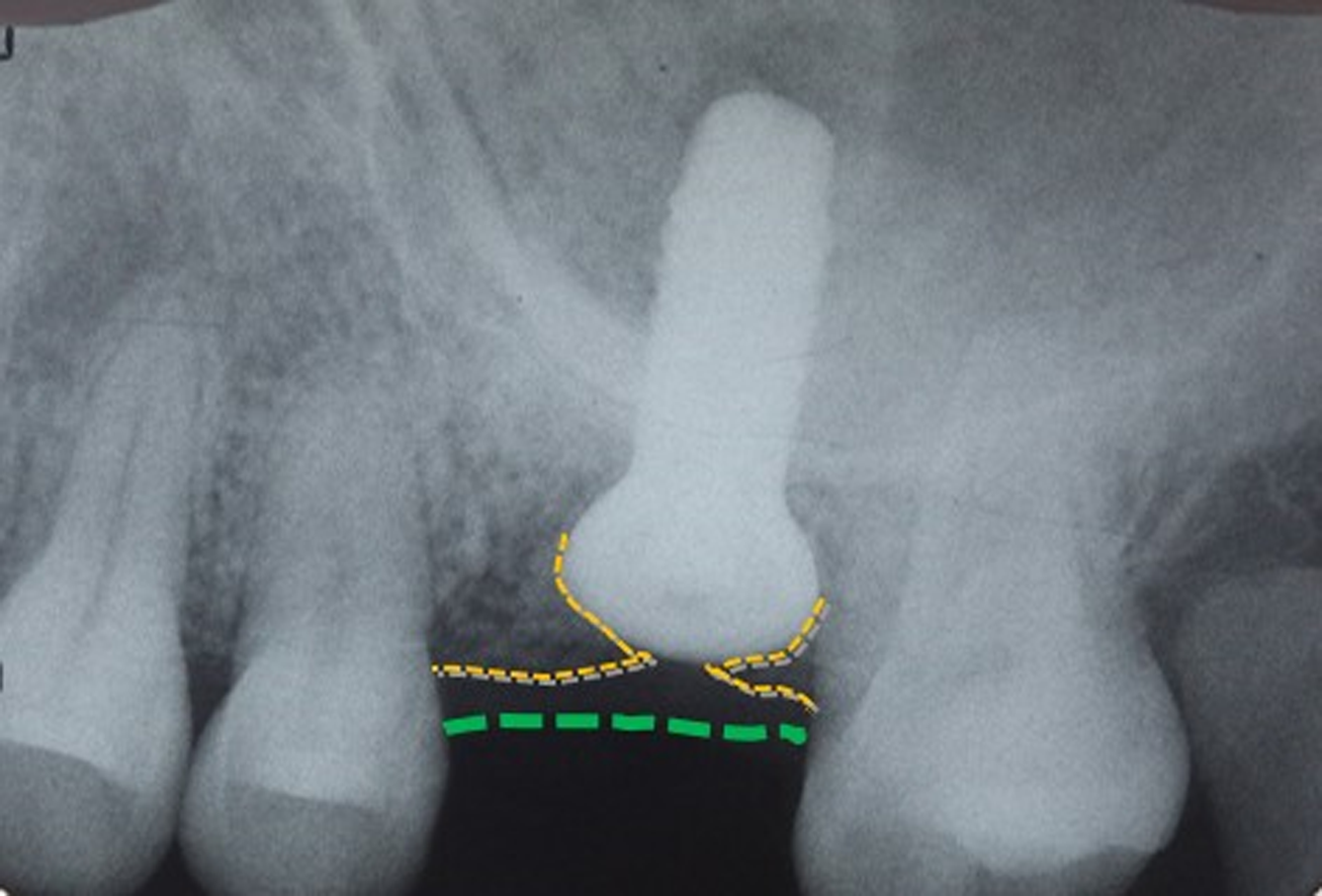
Figure 4 Postoperative periapical standard radiograph in which the regeneration of bone tissue and soft tissue is visible.
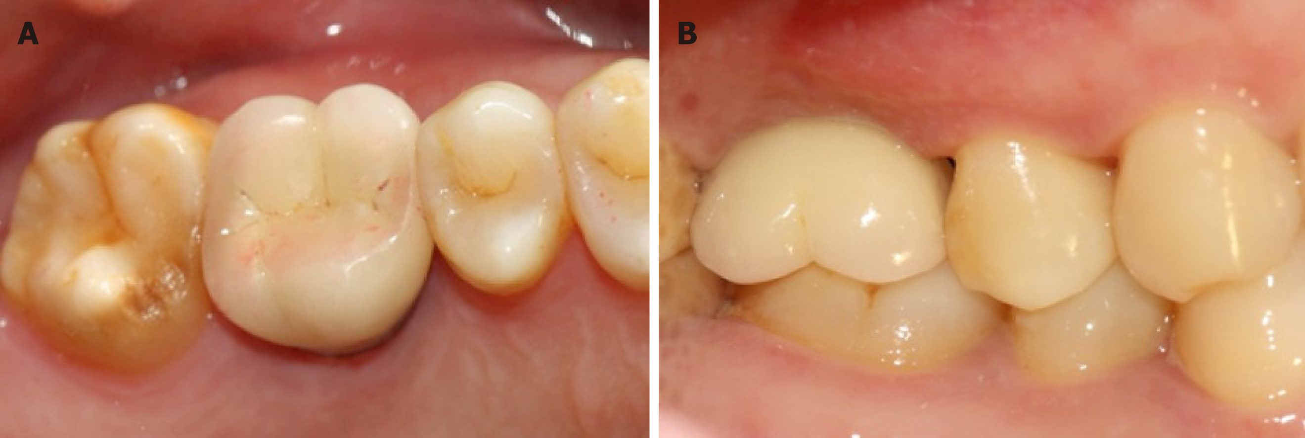
Figure 5 The definitive restoration.
A: Occlusal view; B: Buccal view.
- Citation: Sun XL, Mudalal M, Qi ML, Sun Y, Du LY, Wang ZQ, Zhou YM. Flapless immediate implant placement into fresh molar extraction socket using platelet-rich fibrin: A case report. World J Clin Cases 2019; 7(19): 3153-3159
- URL: https://www.wjgnet.com/2307-8960/full/v7/i19/3153.htm
- DOI: https://dx.doi.org/10.12998/wjcc.v7.i19.3153


