Copyright
©The Author(s) 2019.
World J Clin Cases. Oct 6, 2019; 7(19): 3104-3110
Published online Oct 6, 2019. doi: 10.12998/wjcc.v7.i19.3104
Published online Oct 6, 2019. doi: 10.12998/wjcc.v7.i19.3104
Figure 1 Type I neurofibromatosis patient with right abdominal mass puncture.
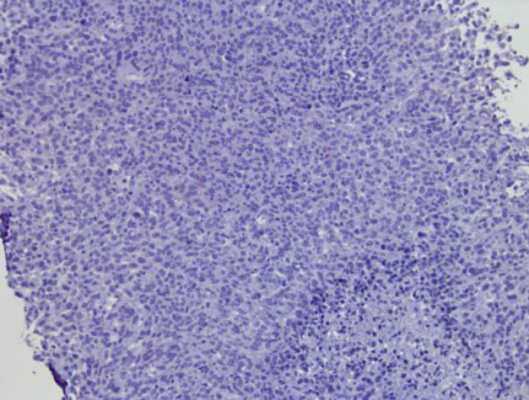
Figure 2 Immunohistochemistry results (programmed death-1 ligand negative).
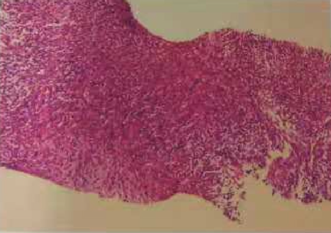
Biopsy pathology image (hematoxylin-eosin staining).
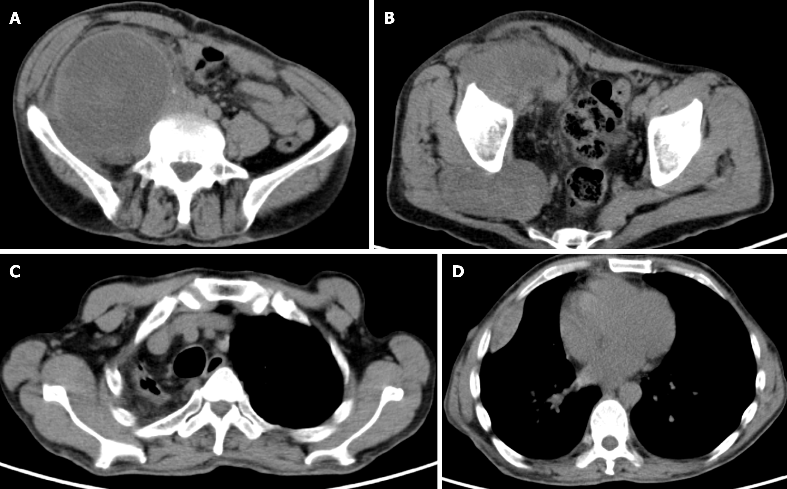
Figure 3 Positron emission tomography/computed tomography image of type I neurofibromatosis patient.
A: Soft tissue masses in the subpleural and horizontal fissure of the upper middle lobe of the right lung. Multiple soft tissue nodules were observed subcutaneously on both chest walls. Mediastinum and main bronchus shift to the right; B: Multiple adjacent spindle shapes were seen in the lateral pleura of the middle lobe of right lung, and fluorodeoxyglucose metabolism was increased in the peripheral area; C: In the right axilla, a large soft tissue mass is seen. The size is about 11.1 cm × 9.3 cm × 17.5 cm, the metabolism of fluorodeoxyglucose is increased, the upper part showed uneven cystic changes, and the cystic area was irradiated with relatively low uptake; the right diaphragm is involved; D: The soft tissue mass in front of the right gluteus maximus. The mass is 8.9 cm × 5.1 cm, and the metabolism of fluorodeoxyglucose is increased.
- Citation: Zhang Y, Chao JJ, Liu XF, Qin SK. Type I neurofibromatosis with spindle cell sarcoma: A case report. World J Clin Cases 2019; 7(19): 3104-3110
- URL: https://www.wjgnet.com/2307-8960/full/v7/i19/3104.htm
- DOI: https://dx.doi.org/10.12998/wjcc.v7.i19.3104


