Copyright
©The Author(s) 2019.
World J Clin Cases. Oct 6, 2019; 7(19): 3090-3097
Published online Oct 6, 2019. doi: 10.12998/wjcc.v7.i19.3090
Published online Oct 6, 2019. doi: 10.12998/wjcc.v7.i19.3090
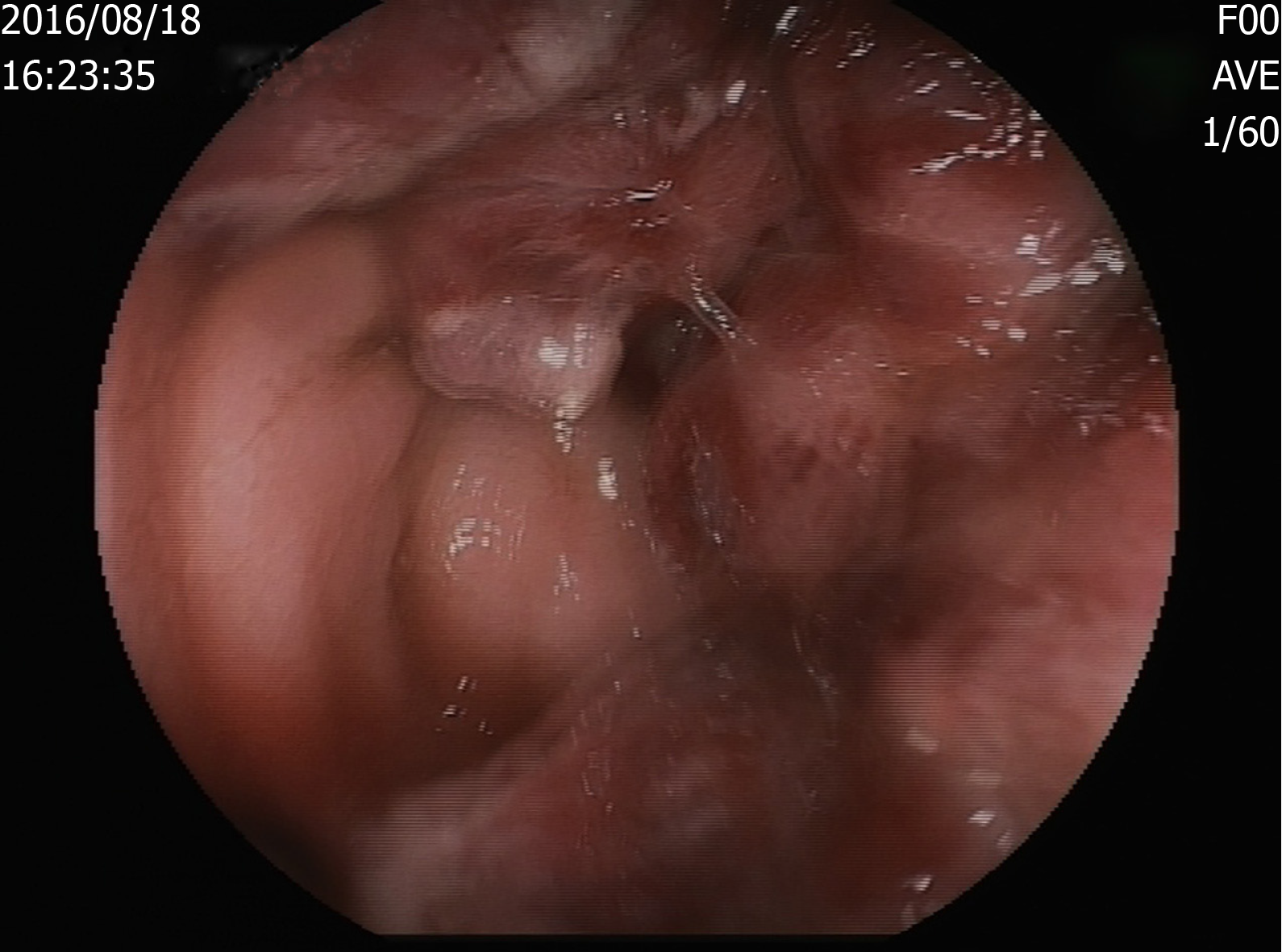
Figure 1 Soft mass on the rectum wall.
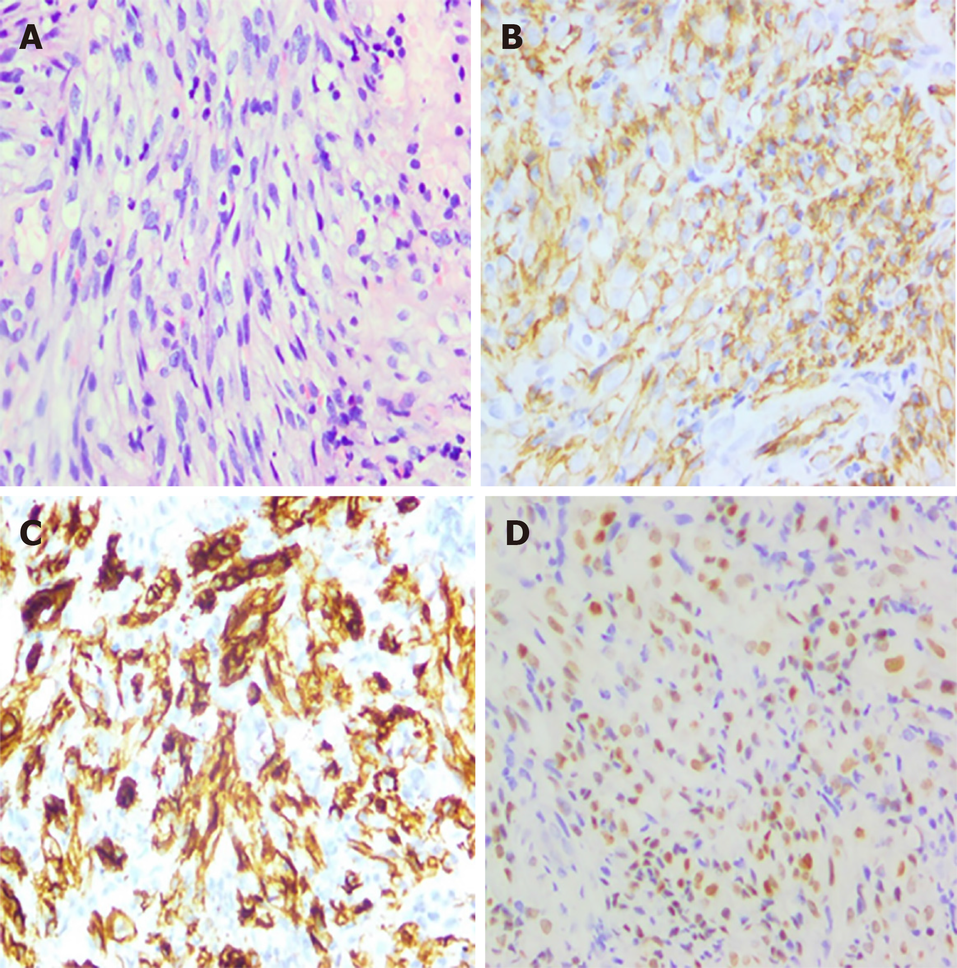
Figure 2 Haematoxylin and eosin stain and immunohistochemistry.
A: Proliferating spindle cells with severe atypia and a certain amount of inflammatory cells scattering among spindle cells (× 200); B: Immunohistochemical stains: positive for CD31, revealing the rectum with KS (× 200); C: Immunohistochemical stains: positive for CD34, revealing the rectum with KS (× 200); D: Immunohistochemical stains: positive for HHV8 antigen (× 200).
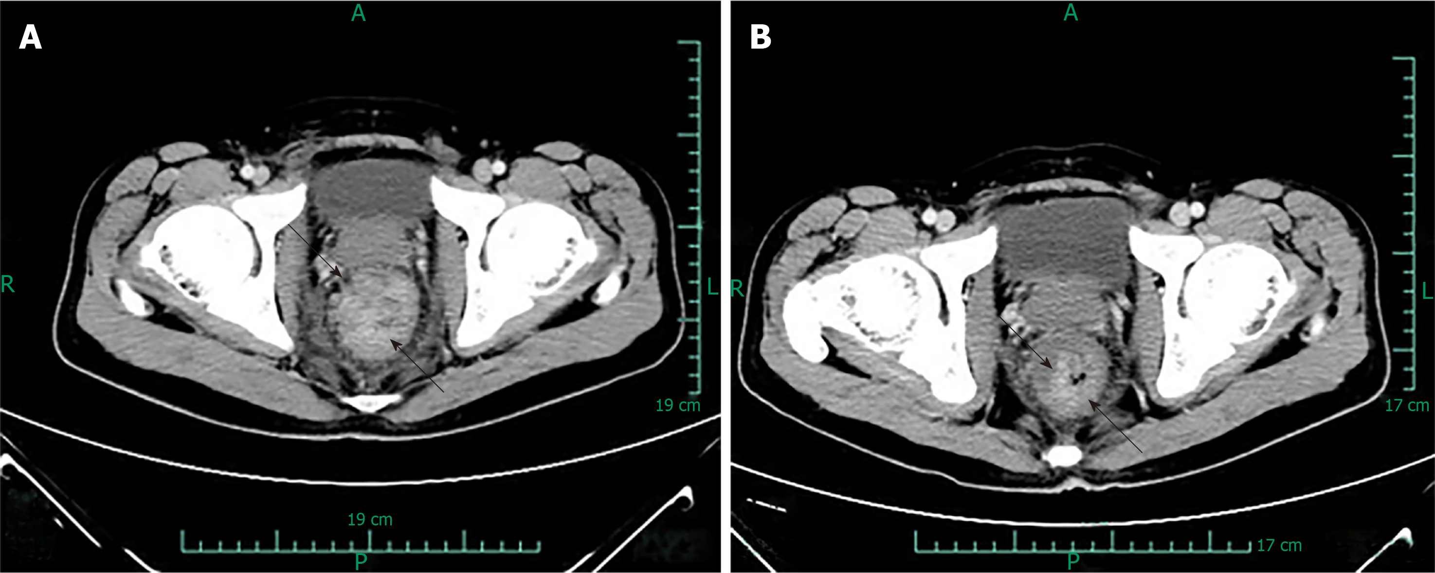
Figure 3 Abdominal CT images of KS.
These two figures are patient’s abdominal CT results scanned before and after chemotherapy. The distance between two arrows is the size of the tumour. A: The size was 4.98 cm and was scanned before chemotherapy on August 16, 2016, which showed thickened wall in the middle-lower rectum; B: The size was 4.18 cm and was scanned after four rounds of chemotherapy on November 21, 2016, which showed that the focus had shrunk.
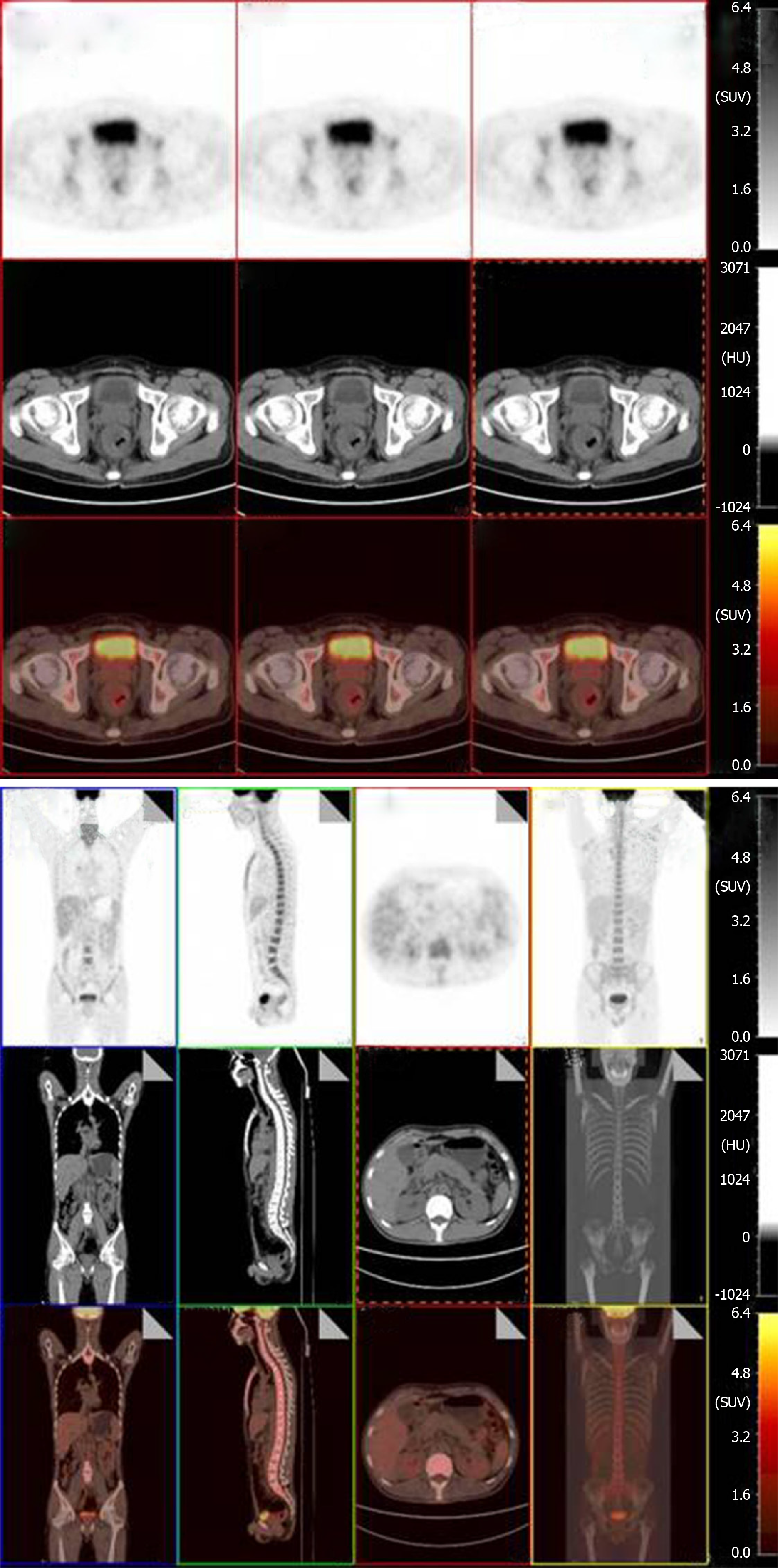
Figure 4 Positron emission tomography images of the Kaposi’s sarcoma.
Positron emission tomography-computed tomography confirmed post chemotherapy of Kaposi’s sarcoma, which showed great improvement in hypermetabolic activity in the rectum with mild residual activity.
- Citation: Zhou QH, Guo YZ, Dai XH, Zhu B. Kaposi’s sarcoma manifested as lower gastrointestinal bleeding in a HIV/HBV-co-infected liver cirrhosis patient: A case report. World J Clin Cases 2019; 7(19): 3090-3097
- URL: https://www.wjgnet.com/2307-8960/full/v7/i19/3090.htm
- DOI: https://dx.doi.org/10.12998/wjcc.v7.i19.3090


