Copyright
©The Author(s) 2019.
World J Clin Cases. Oct 6, 2019; 7(19): 3082-3089
Published online Oct 6, 2019. doi: 10.12998/wjcc.v7.i19.3082
Published online Oct 6, 2019. doi: 10.12998/wjcc.v7.i19.3082
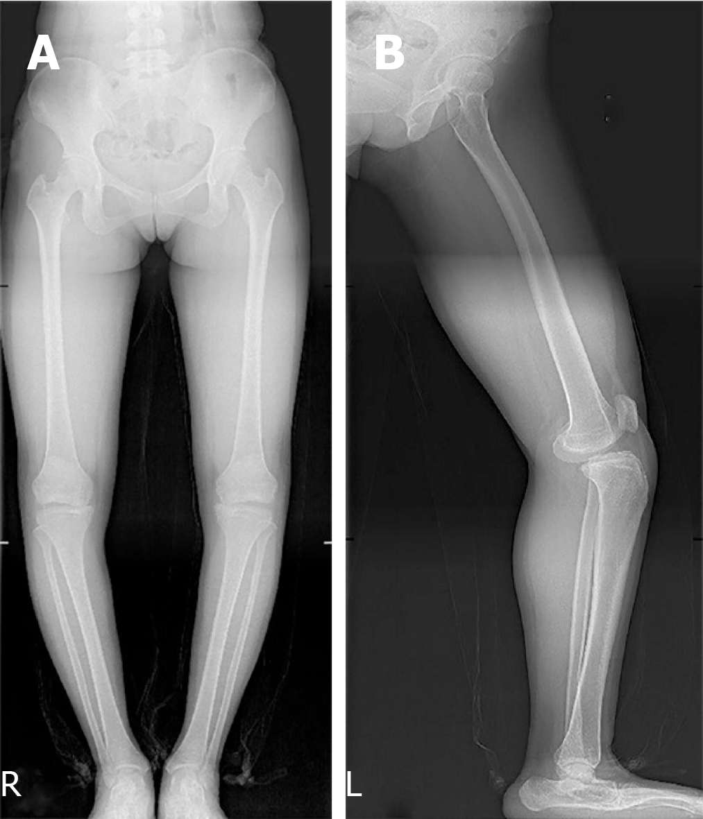
Figure 1 X-ray images.
A: Unequal width of the joint spaces; B: Anterior and medial displacement of the tibial plate.
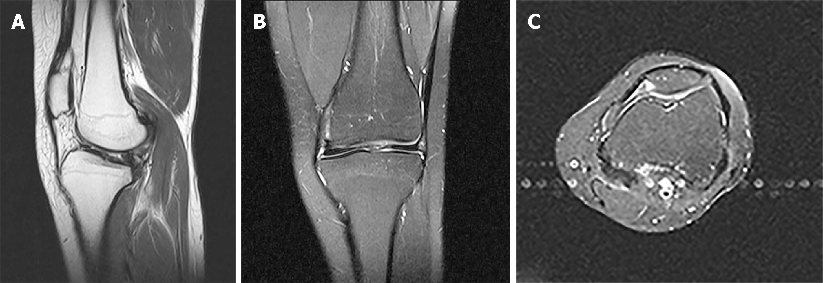
Figure 2 Magnetic resonance imaging scan showing cruciate ligament deficiency.
A: Left knee joint anterior displacement with subluxation; B: Mild abrasion in the medial tibial platform; C: Femoral articular surface wear.
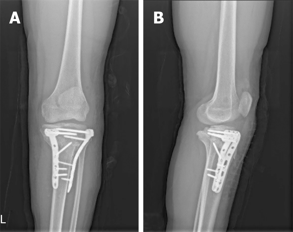
Figure 3 X-ray images showing the left knee with no loosening of the internal fixation.
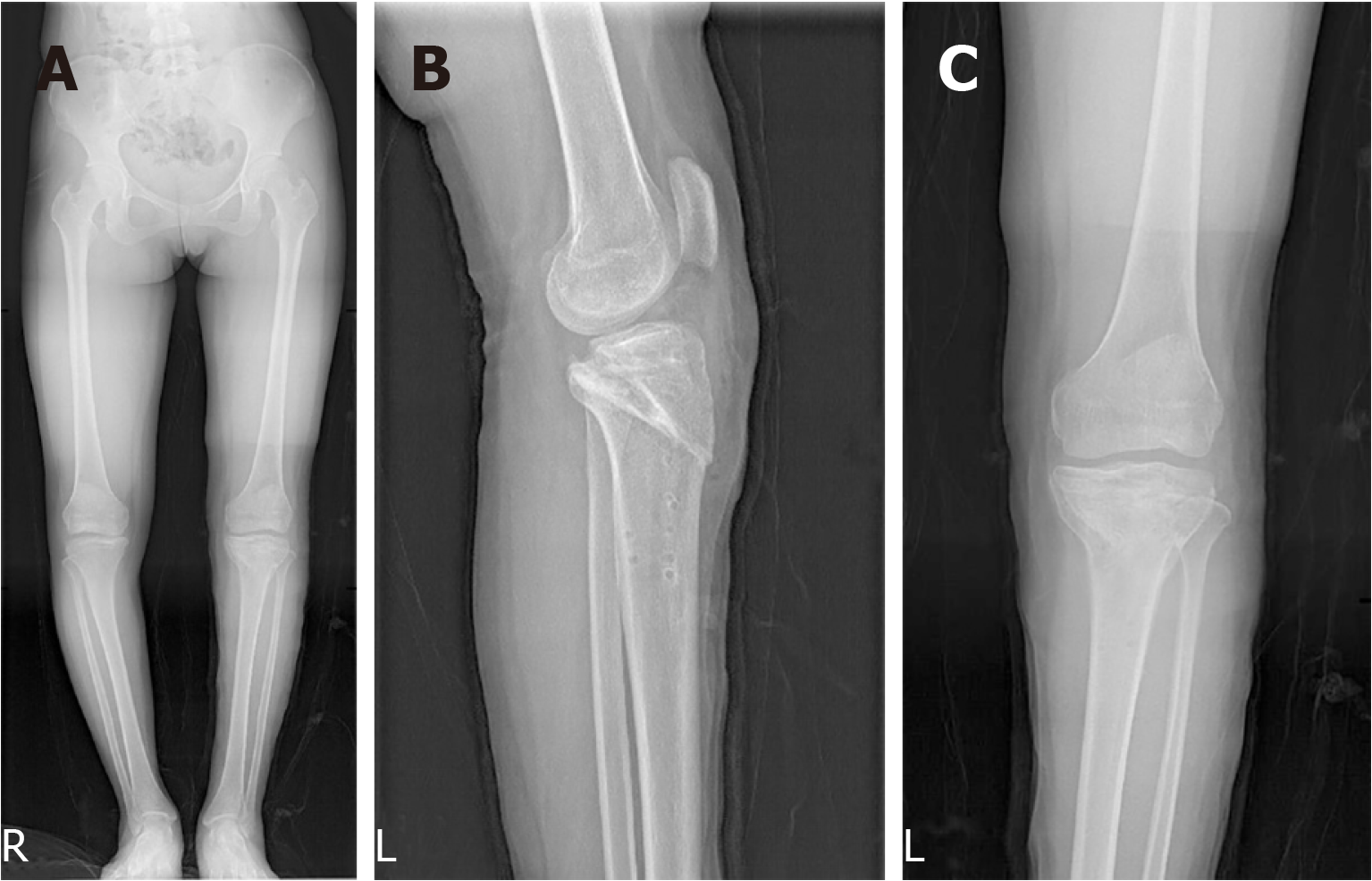
Figure 4 X-ray images.
A: Correct alignment of the left knee and unequal length of the lower limbs with right knee inversion to the medial; B: Left knee tibial plateaus moving inwards; C: Equal width of the joint spaces.
- Citation: Lu R, Zhu DP, Chen N, Sun H, Li ZH, Cao XW. How should congenital absence of cruciate ligaments be treated? A case report and literature review. World J Clin Cases 2019; 7(19): 3082-3089
- URL: https://www.wjgnet.com/2307-8960/full/v7/i19/3082.htm
- DOI: https://dx.doi.org/10.12998/wjcc.v7.i19.3082


