Copyright
©The Author(s) 2019.
World J Clin Cases. Oct 6, 2019; 7(19): 3055-3061
Published online Oct 6, 2019. doi: 10.12998/wjcc.v7.i19.3055
Published online Oct 6, 2019. doi: 10.12998/wjcc.v7.i19.3055
Figure 1 First renal biopsy manifestations.
A: Light microscopy showed mild increased mesangial matrix and mesangial hypercellularity (periodic acid-Schiff-methenamine stain, 200 ×); B: Immunofluorescence showed deposits of IgA in the mesangium (200 ×); C: Electron microscopy showed electron-dense deposits in the mesangium (blue arrow).
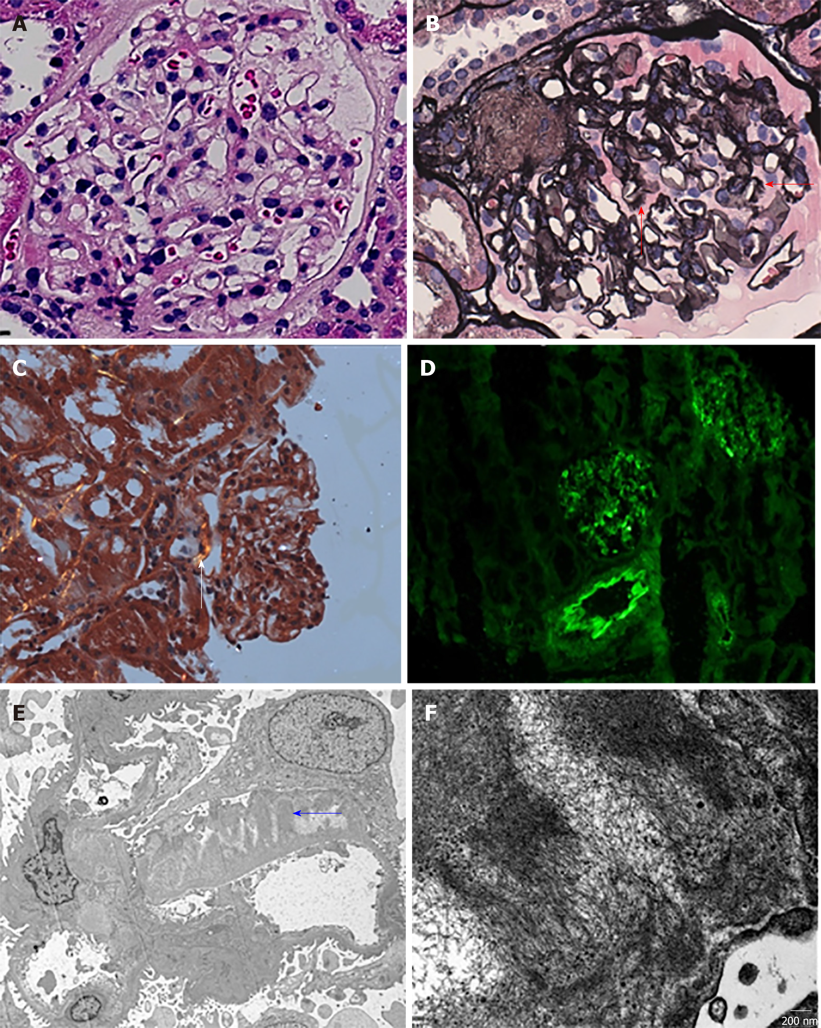
Figure 2 Second renal biopsy manifestations.
A: Light microscopy showed well-opened capillary loops and mild pale eosinophilic material in the mesangium and basement membranes (hematoxylin and eosin stain, 400 ×); B: Thickened GBM with subepithelial fringe-like projections (red arrow) (periodic acid-Schiff-methenamine stain, 400 ×); C: Congo red stain was greenish under polarized light (white arrow, 100 ×), involving an afferent glomerular arteriole; D: Immunofluorescence showed deposits of IgM in the mesangium and small vessels (200 ×). κ and λ were also positive (not shown); E and F: Electron microscopy showed randomly oriented amyloid fibrils along glomerular capillary walls (blue arrow).
Figure 3 Flow chart.
- Citation: Wu HT, Wen YB, Ye W, Liu BY, Shen KN, Gao RT, Li MX. Underlying IgM heavy chain amyloidosis in treatment-refractory IgA nephropathy: A case report. World J Clin Cases 2019; 7(19): 3055-3061
- URL: https://www.wjgnet.com/2307-8960/full/v7/i19/3055.htm
- DOI: https://dx.doi.org/10.12998/wjcc.v7.i19.3055


