Copyright
©The Author(s) 2019.
World J Clin Cases. Oct 6, 2019; 7(19): 3047-3054
Published online Oct 6, 2019. doi: 10.12998/wjcc.v7.i19.3047
Published online Oct 6, 2019. doi: 10.12998/wjcc.v7.i19.3047
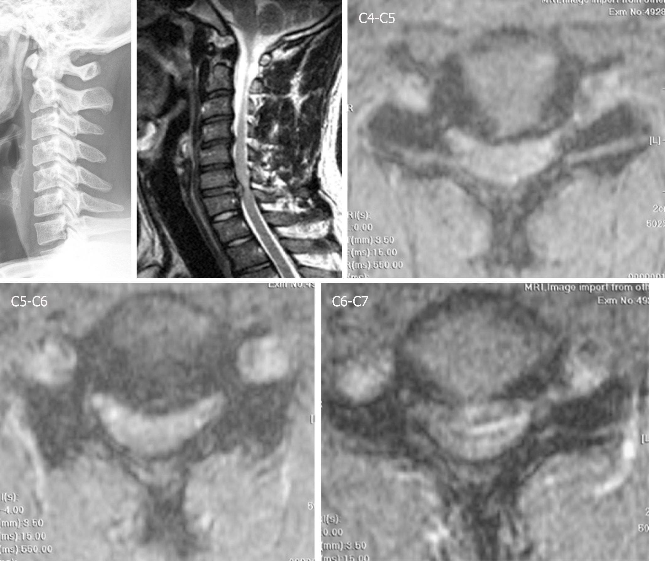
Figure 1 Herniation of the intervertebral disc at the C4-C5, C5-C6, and C6-C7 levels.
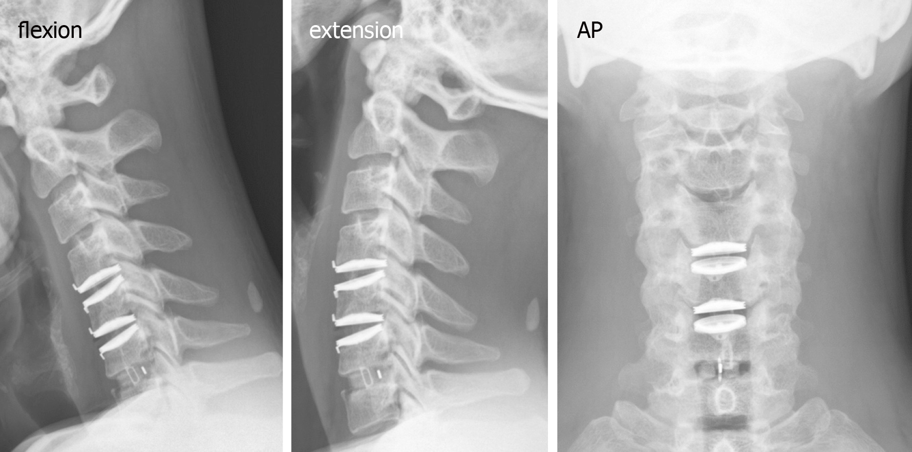
Figure 2 Postoperative cervical radiographs in the dynamic (flexion and extension) and anteroposterior views.
AP: Anteroposterior.
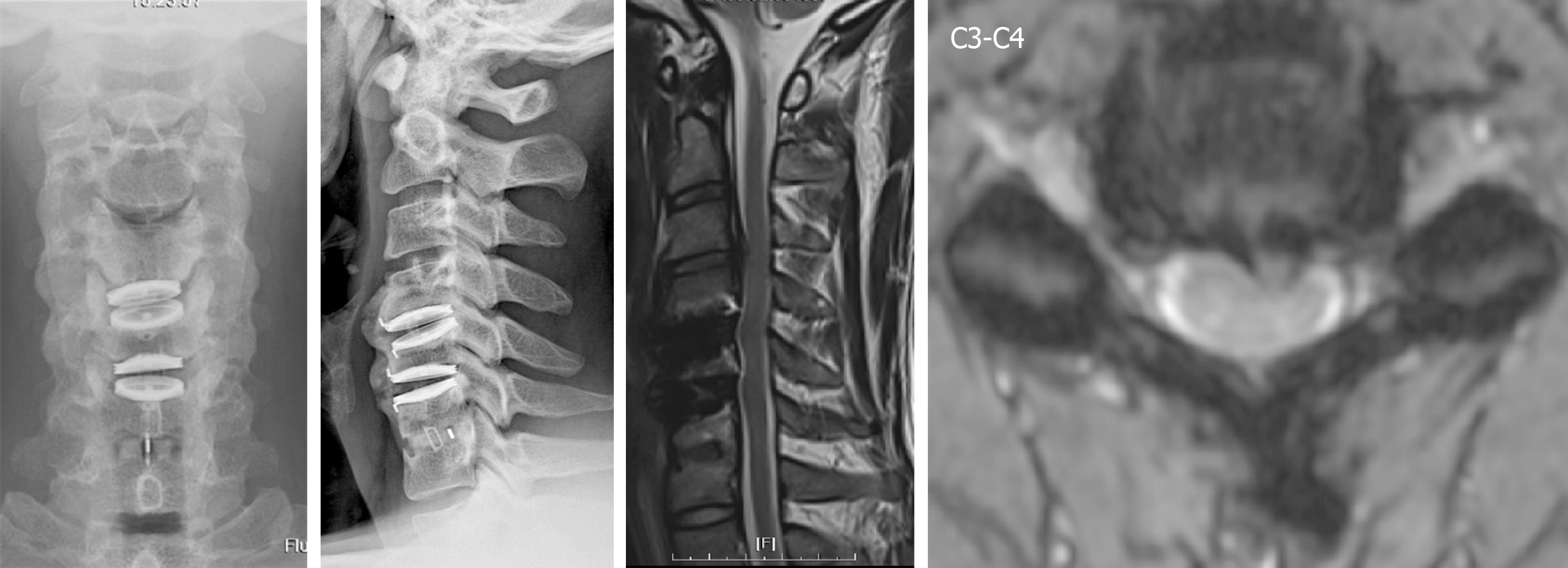
Figure 3 C3-C4 herniation of the intervertebral disc with spinal cord compression in the fourth year after the operation.
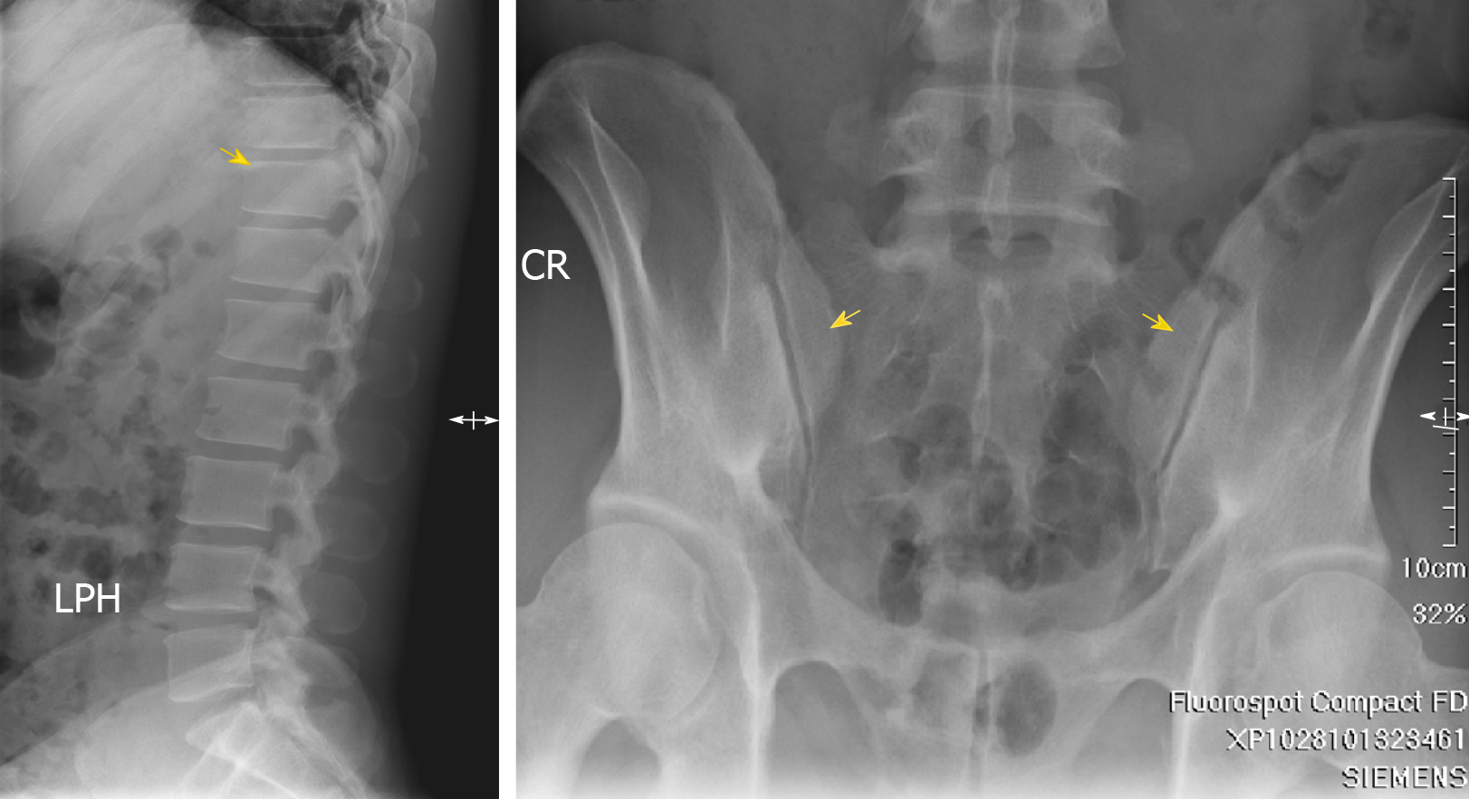
Figure 4 Initial X-rays at four years after the motorcycle accident.
Left: Shiny corner sign, a sclerotic margin of the T-L vertebral body corner, compatible with seronegative spondyloarthropathy; Right: Bone sclerotic change in the bilateral sacroiliac joint, compatible with bilateral sacroiliitis, grade I.
Figure 5 Pre-operative and postoperative radiographs during follow-up.
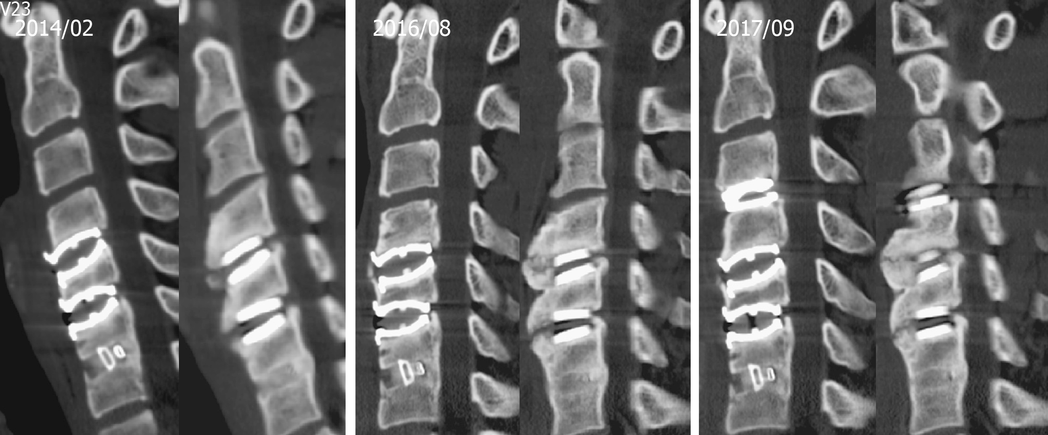
Figure 6 Cervical computed tomography images showing anterior spur formation without spinal canal stenosis.
February 2014: 1.5 years after the first operation; August 2016: 4 years after the first operation; September 2017: 1 year after the second operation.
- Citation: Huang CW, Tang CL, Pan HC, Tzeng CY, Tsou HK. Severe heterotopic ossification in a seronegative spondyloarthritis patient after cervical Bryan disc arthroplasty: A case report. World J Clin Cases 2019; 7(19): 3047-3054
- URL: https://www.wjgnet.com/2307-8960/full/v7/i19/3047.htm
- DOI: https://dx.doi.org/10.12998/wjcc.v7.i19.3047


