Copyright
©The Author(s) 2019.
World J Clin Cases. Sep 26, 2019; 7(18): 2871-2878
Published online Sep 26, 2019. doi: 10.12998/wjcc.v7.i18.2871
Published online Sep 26, 2019. doi: 10.12998/wjcc.v7.i18.2871
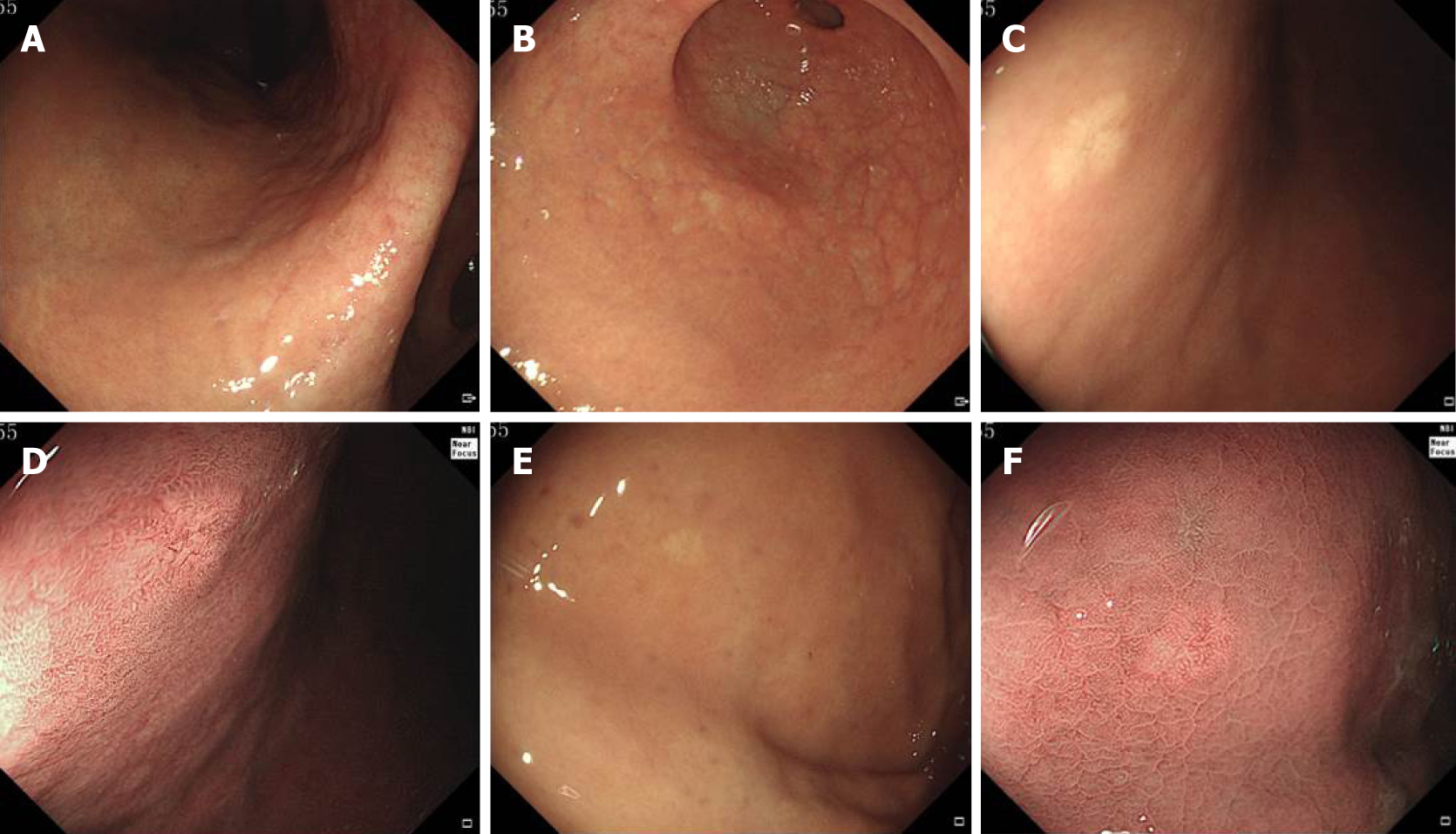
Figure 1 Endoscopic findings of all lesions.
A and B: The gastric mucosa showed grade C-2 atrophic gastritis according to the Kimura-Takemoto classification; C: White light endoscopy revealed a submucosal tumor-like elevated mass with a whitish mucosal surface on the anterior gastric corpus wall; D: Narrow-band imaging showed an irregular microvascular pattern and dilatation of microvessels with branching architecture; E: White light endoscopy revealed a flat shaped lesion with a whitish mucosal surface on the gastric fundus; F: Narrow-band imaging showed regular and dilated microvessels.
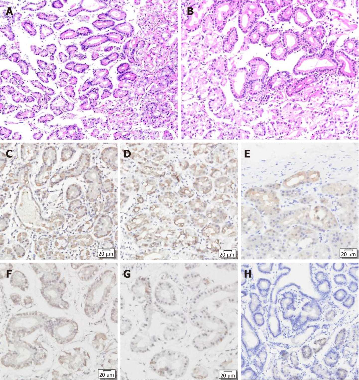
Figure 2 Hematoxylin and eosin staining showed that the tumors had clear demarcation from the surrounding fundus glands and an irregular glandular structure.
The tumors were composed of chief cell-like cells with mild nuclear atypia. A: Gastric corpus; B: Gastric fundus; C-H: Immunohistochemical staining showed that both lesions were positive for pepsinogen I (C and F) and MUC6 (D and G), and partially positive for H+/K+-ATPase (E and H).
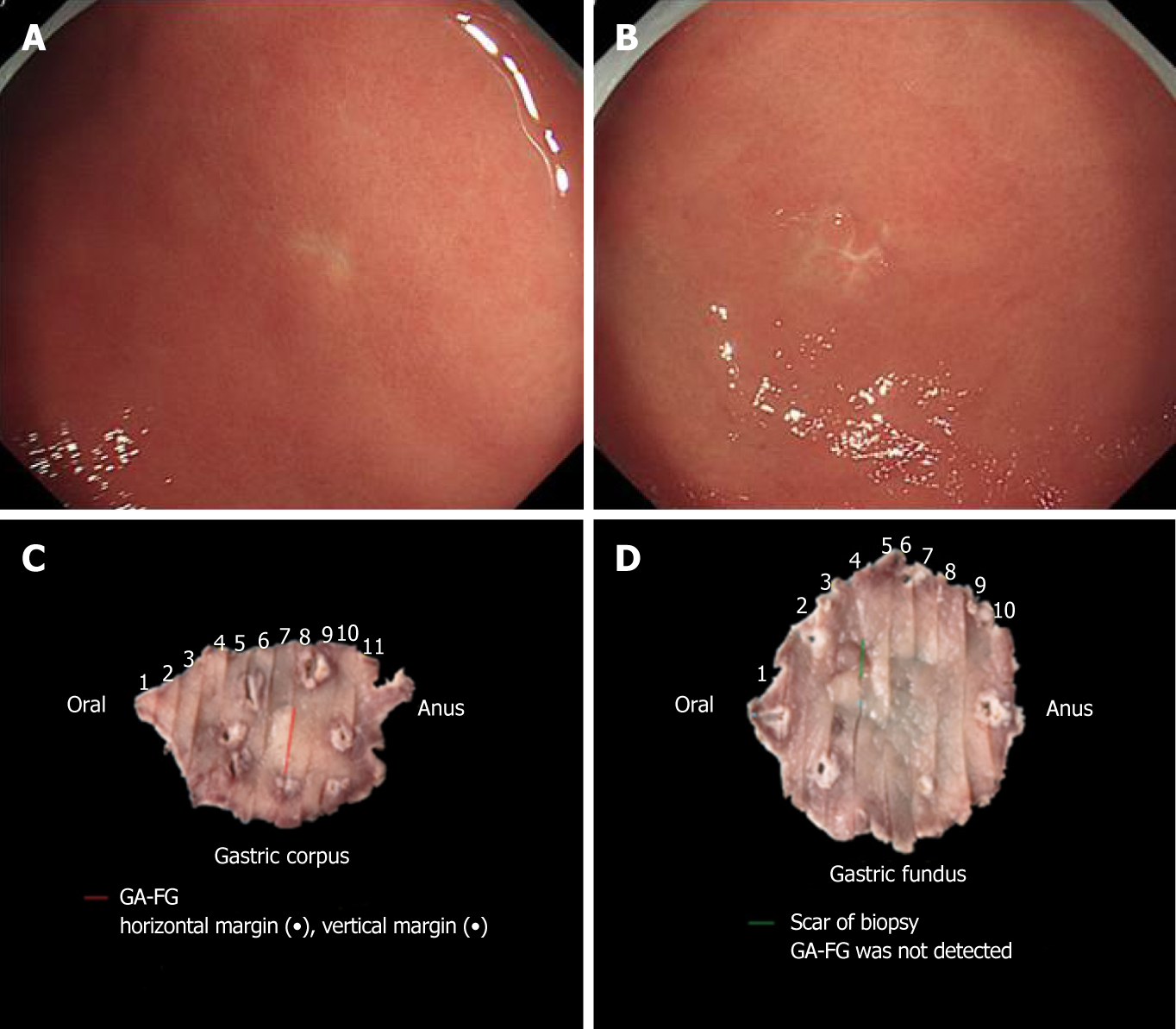
Figure 3 Endoscopic submucosal dissection and pathological reconstruction of the scar from the biopsy.
A and B: White light endoscopy revealed the scar of the biopsy of the gastric corpus (B) and gastric fundus (A) after biopsy; C and D: The endoscopic submucosal dissection-resected specimens of the gastric fundus (C) and gastric corpus (D) (pathological reconstruction).
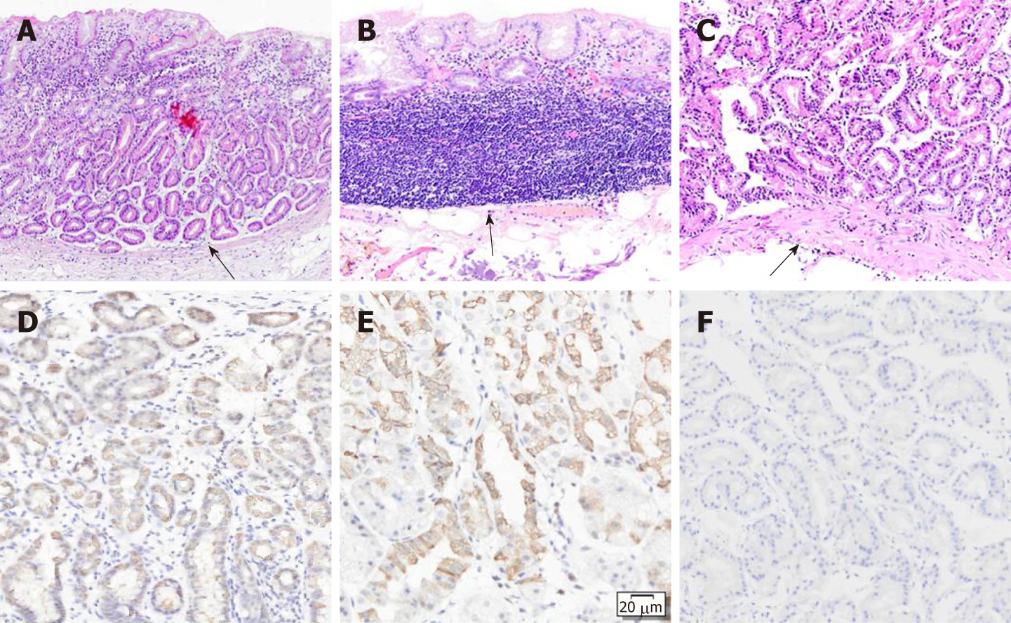
Figure 4 Hematoxylin and eosin staining.
A: Hematoxylin and eosin staining showed that the gastric corpus lesion was located in the deep layer of the lamina propria and did not invade into the muscularis mucosa. The lesion did not extend to the superficial epithelium; B: Hematoxylin and eosin staining showed no detectable lesion in the gastric fundus but only the biopsy scar. The mucularis mucosa of the ESD-resected specimen was intact; C: Hematoxylin and eosin staining of the fundic biopsy showing that the mucularis mucosa was intact; D-F: Immunostaining showed that both lesions were positive for pepsinogen I (D) and MUC6 (E) and partially positive for H+/K+-ATPase (F).
- Citation: Chen O, Shao ZY, Qiu X, Zhang GP. Multiple gastric adenocarcinoma of fundic gland type: A case report. World J Clin Cases 2019; 7(18): 2871-2878
- URL: https://www.wjgnet.com/2307-8960/full/v7/i18/2871.htm
- DOI: https://dx.doi.org/10.12998/wjcc.v7.i18.2871


