Copyright
©The Author(s) 2019.
World J Clin Cases. Sep 26, 2019; 7(18): 2838-2842
Published online Sep 26, 2019. doi: 10.12998/wjcc.v7.i18.2838
Published online Sep 26, 2019. doi: 10.12998/wjcc.v7.i18.2838
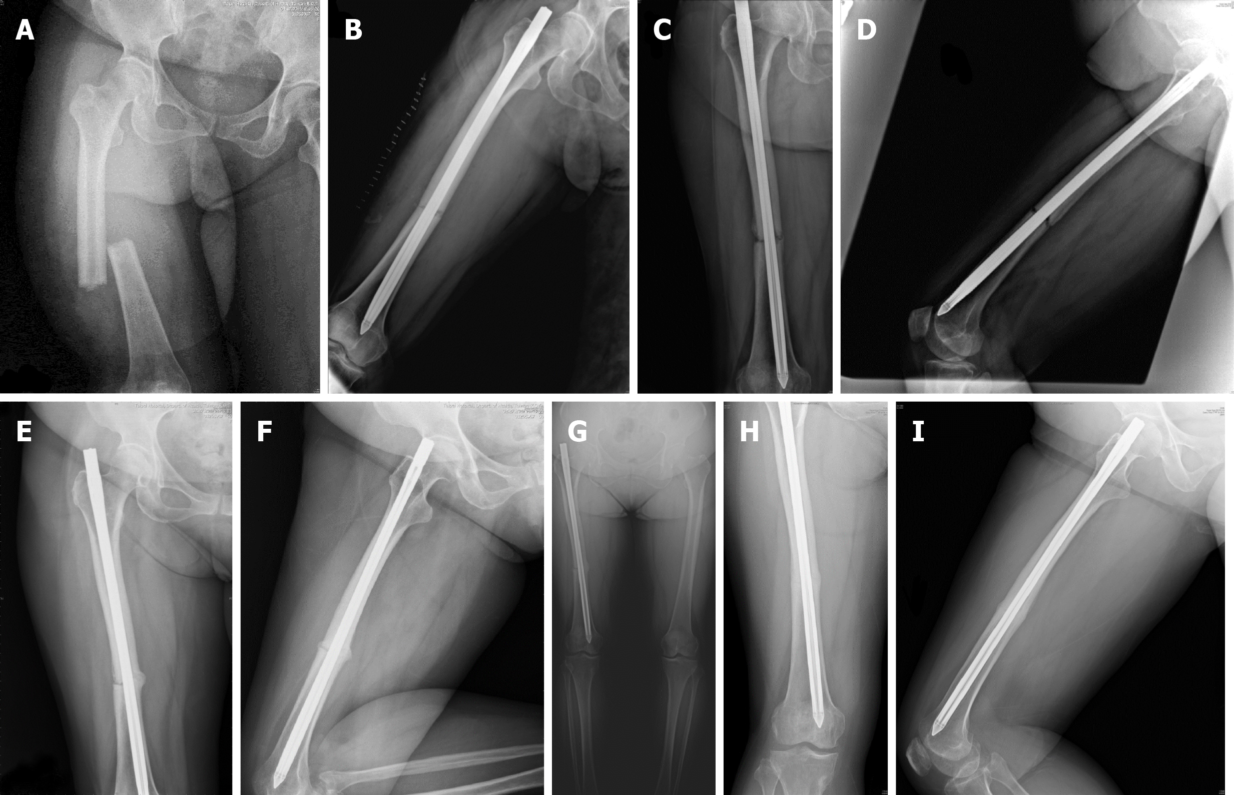
Figure 1 Radiographs of the femur.
A: Anteroposterior view. Right femoral shaft fracture, middle third, simple transverse. Date: April/12/2016; B: Lateral view. Status after the closed reduction internal fixation with the Fixion nail. Date: April/13/2016; C: Anteroposterior view, 6 mo postoperatively, one month before the teriparatide treatment. No callus was observed. Date: Oct/20/2016; D: Lateral view. Date: Oct/20/2016; E: Anteroposterior view, 3 mo after teriparatide use. A callus was observed. Date: Feb/21/2017; F: Lateral view. Date: Feb/21/2017; G: Anteroposterior view, 5 mo after teriparatide use. Continuous improvement in fracture gap reduction and bone bridging was observed. Date: April/21/2017; H: Anteroposterior view, 6 mo after the discontinuation of teriparatide. A complete union was observed. Date: Nov/14/2017; I: Lateral view. Date: Nov/14/2017.
- Citation: Tsai MH, Hu CC. Teriparatide as nonoperative treatment for femoral shaft atrophic nonunion: A case report. World J Clin Cases 2019; 7(18): 2838-2842
- URL: https://www.wjgnet.com/2307-8960/full/v7/i18/2838.htm
- DOI: https://dx.doi.org/10.12998/wjcc.v7.i18.2838


