Copyright
©The Author(s) 2019.
World J Clin Cases. Sep 26, 2019; 7(18): 2823-2830
Published online Sep 26, 2019. doi: 10.12998/wjcc.v7.i18.2823
Published online Sep 26, 2019. doi: 10.12998/wjcc.v7.i18.2823
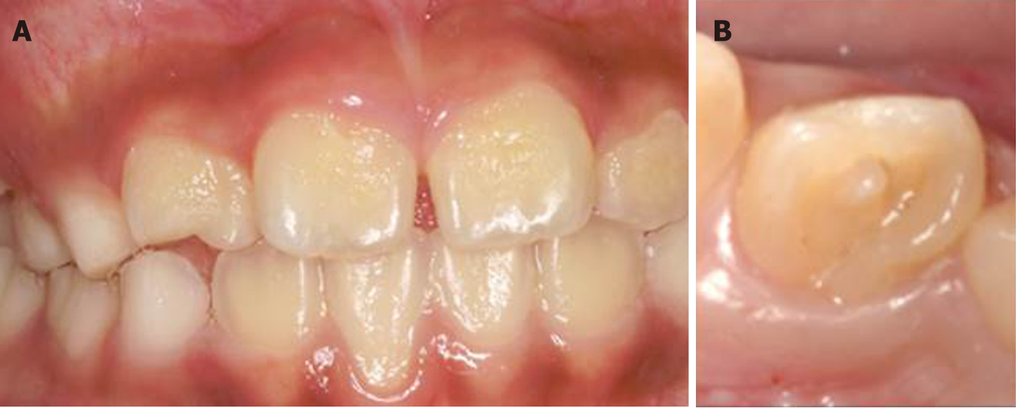
Figure 1 Clinical examination.
A: The intraoral examination revealed normal soft tissue; B: Occlusal view shows a talon cusp and square-shaped crown.
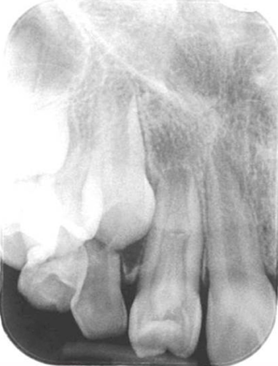
Figure 2 Periapical radiograph.
The image shows a large elliptical space inside the invagination, and invagination of the dentin lined by enamel into the main canal space spreading into the apical part of the tooth root was observed. Tooth 12 appears to be associated with a large periapical radiolucent lesion around an immature root apex.
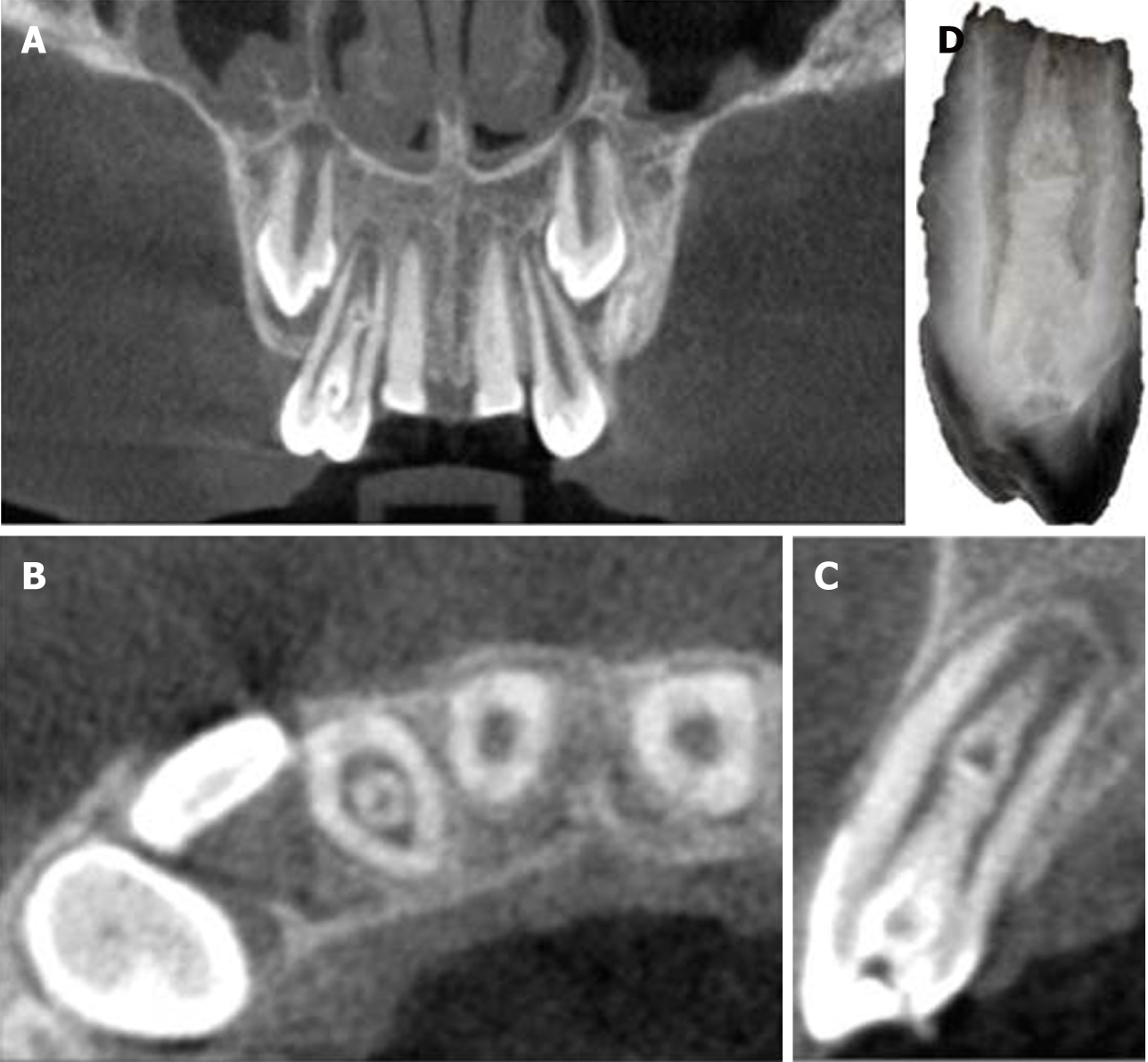
Figure 3 Cone-beam computed tomography images.
A: Coronal view shows a central bulge; B: Axial view shows the relative positions of the main root canal and pseudo root canal; C: Sagittal plane view indicates that the enlargement is connected with the main root canal; D: Reconstruction of the three-dimensional structure of the dens invaginatus.
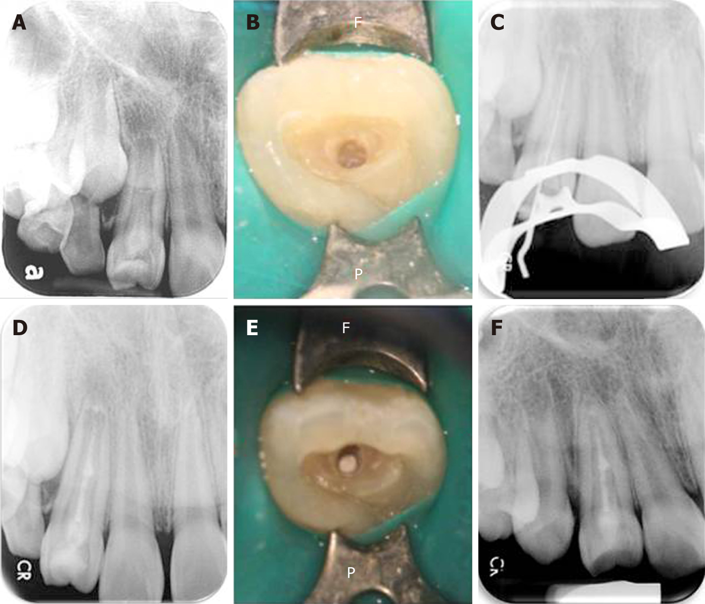
Figure 4 The operating process.
A: Pre-operative radiograph showing the presence of dens invaginatus associated with an open apex and a large periapical lesion. The middle part of the bulge was suspected to be connected to the main root canal; B: The position of the invagination was located under a microscope and access-opening was performed; C: A root ZX mini apex locator and a periapical radiograph were used to determine the working length before conducting complete debridement; D: Considering the interconnections between the root canals in the middle segment, a mineral trioxide aggregate (MTA) sealer was used to fill and seal the pseudo root canal and perform pulp capping in the main canal; E: The MTA filling as observed under the microscope; F: At the one-year follow-up, the apical lesion had healed well (the tooth wall had thickened and the apical foramen was closed). The tooth still responded normally to the pulp vitality test.
- Citation: Lee HN, Chen YK, Chen CH, Huang CY, Su YH, Huang YW, Chuang FH. Conservative pulp treatment for Oehlers type III dens invaginatus: A case report. World J Clin Cases 2019; 7(18): 2823-2830
- URL: https://www.wjgnet.com/2307-8960/full/v7/i18/2823.htm
- DOI: https://dx.doi.org/10.12998/wjcc.v7.i18.2823


