Copyright
©The Author(s) 2019.
World J Clin Cases. Sep 26, 2019; 7(18): 2787-2793
Published online Sep 26, 2019. doi: 10.12998/wjcc.v7.i18.2787
Published online Sep 26, 2019. doi: 10.12998/wjcc.v7.i18.2787
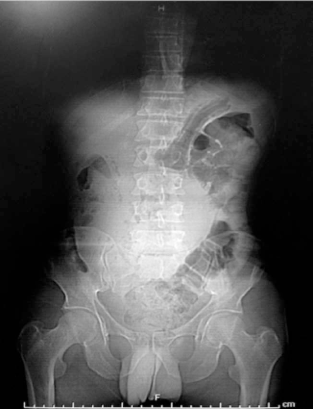
Figure 1 Abdominal radiography with radiopaque area in mesogastrium.
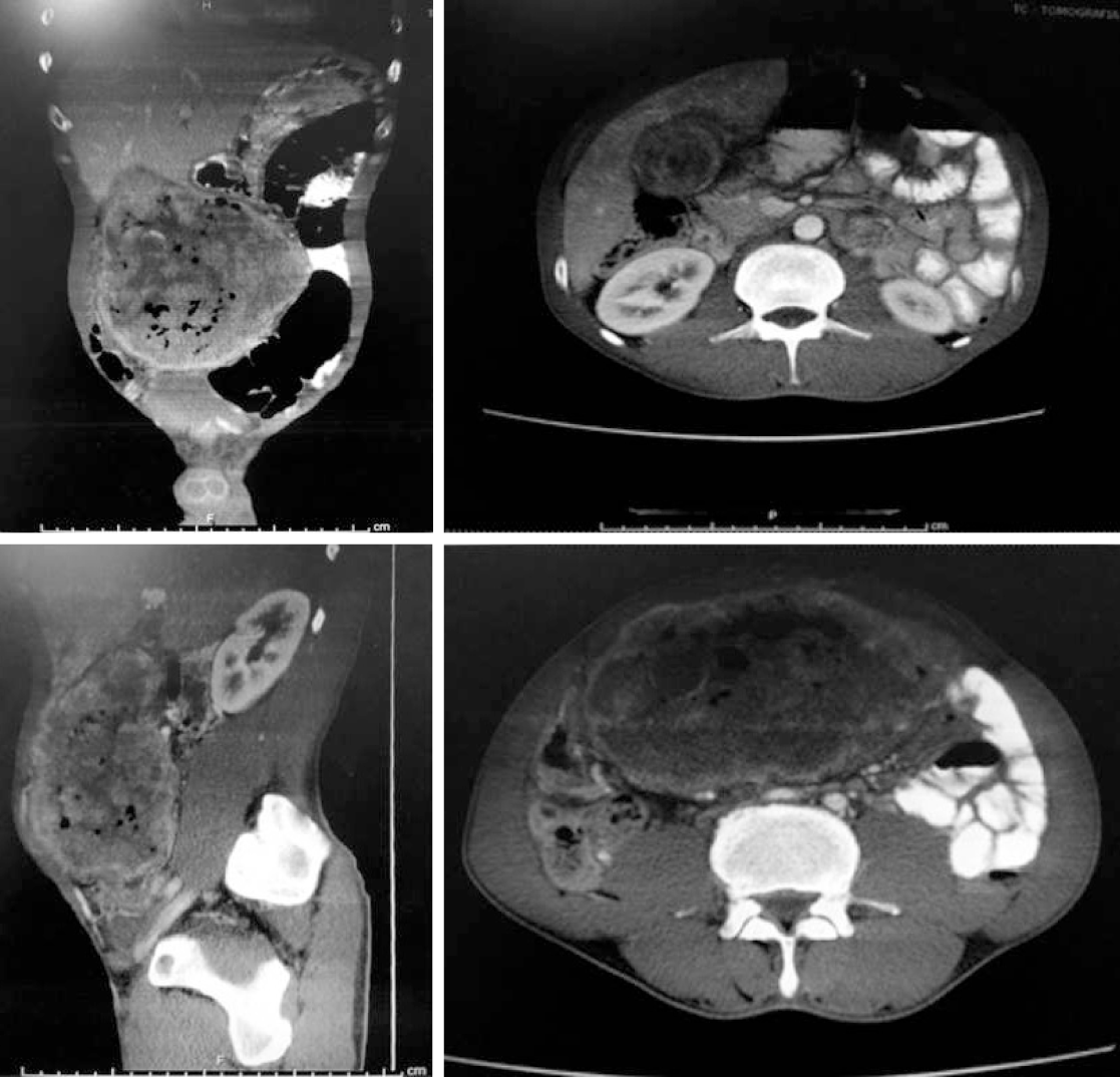
Figure 2 Computed tomography with a coronal, sagittal, and axial image showing a large tumor in the hepatic bed.
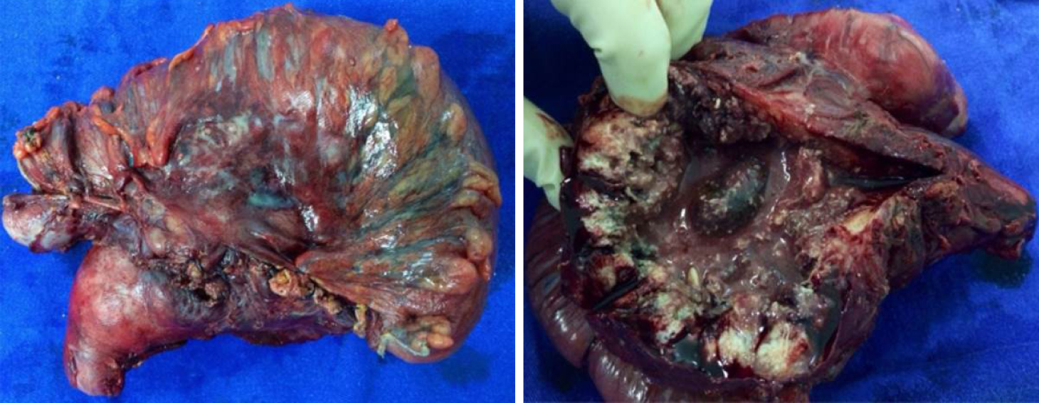
Figure 3 Surgical specimen with en bloc resection of gallbladder and transverse colon segment.
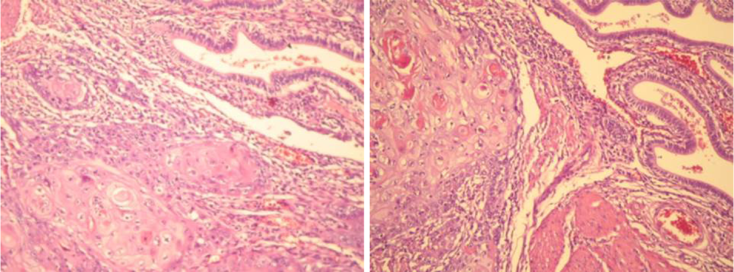
Figure 4 Transition from spinocellular carcinoma to extensive blocks with formation of horny pearls, and the gallbladder mucosa presenting the columnar epithelium.
Hematoxylin-eosin staining, 100 ×.
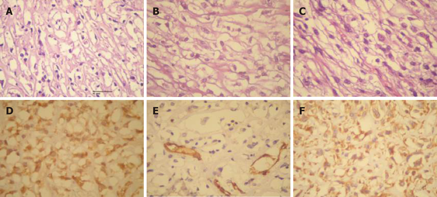
Figure 5 Plexiform fibromyxoma of the gastric wall.
A, B, C: The image shows a fusocellular neoplasm with elongate or oval nuclei and clear cytoplasm with basophilic base of myxoid aspect. Hematoxylin-eosin staining, 400 ×; D: Immunoexpression of actin antibody of smooth muscle in neoplastic cells; E: Immunoexpression of CD31 antibody in blood vessel walls; F: Immunoexpression of vimentin in neoplastic cells.
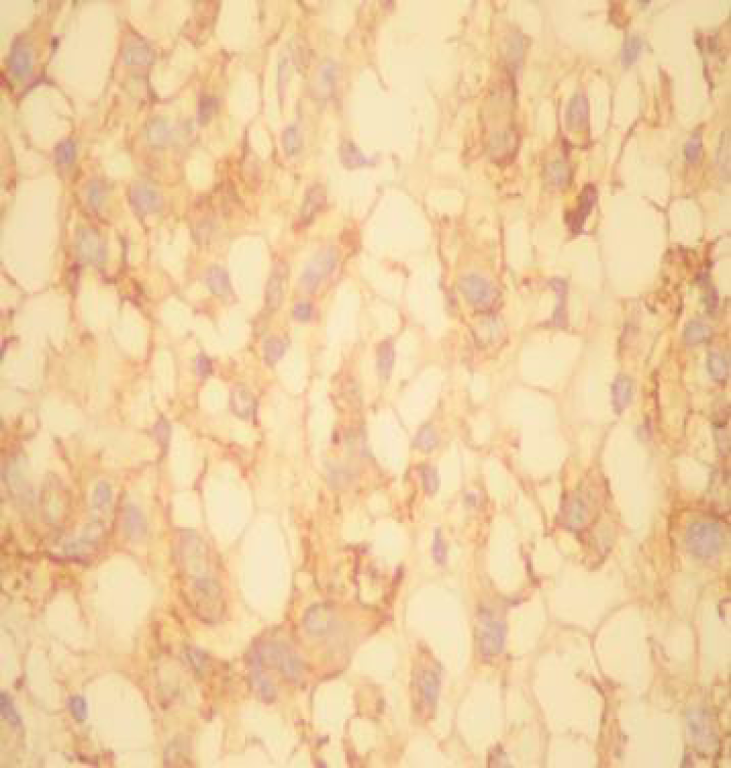
Figure 6 Immunoexpression of CD56 antibody and anti-neuron specify enolase antibody in neoplastic cells.
Absence of immunoexpression of CD117 antibody. Positivity of KI-67 to the nuclei of the neoplastic cells (low index of mitotic proliferation).
- Citation: Junior MAR, Favaro ML, Santin S, Silva CM, Iamarino APM. Giant squamous cell carcinoma of the gallbladder: A case report. World J Clin Cases 2019; 7(18): 2787-2793
- URL: https://www.wjgnet.com/2307-8960/full/v7/i18/2787.htm
- DOI: https://dx.doi.org/10.12998/wjcc.v7.i18.2787


