Copyright
©The Author(s) 2019.
World J Clin Cases. Sep 6, 2019; 7(17): 2652-2657
Published online Sep 6, 2019. doi: 10.12998/wjcc.v7.i17.2652
Published online Sep 6, 2019. doi: 10.12998/wjcc.v7.i17.2652
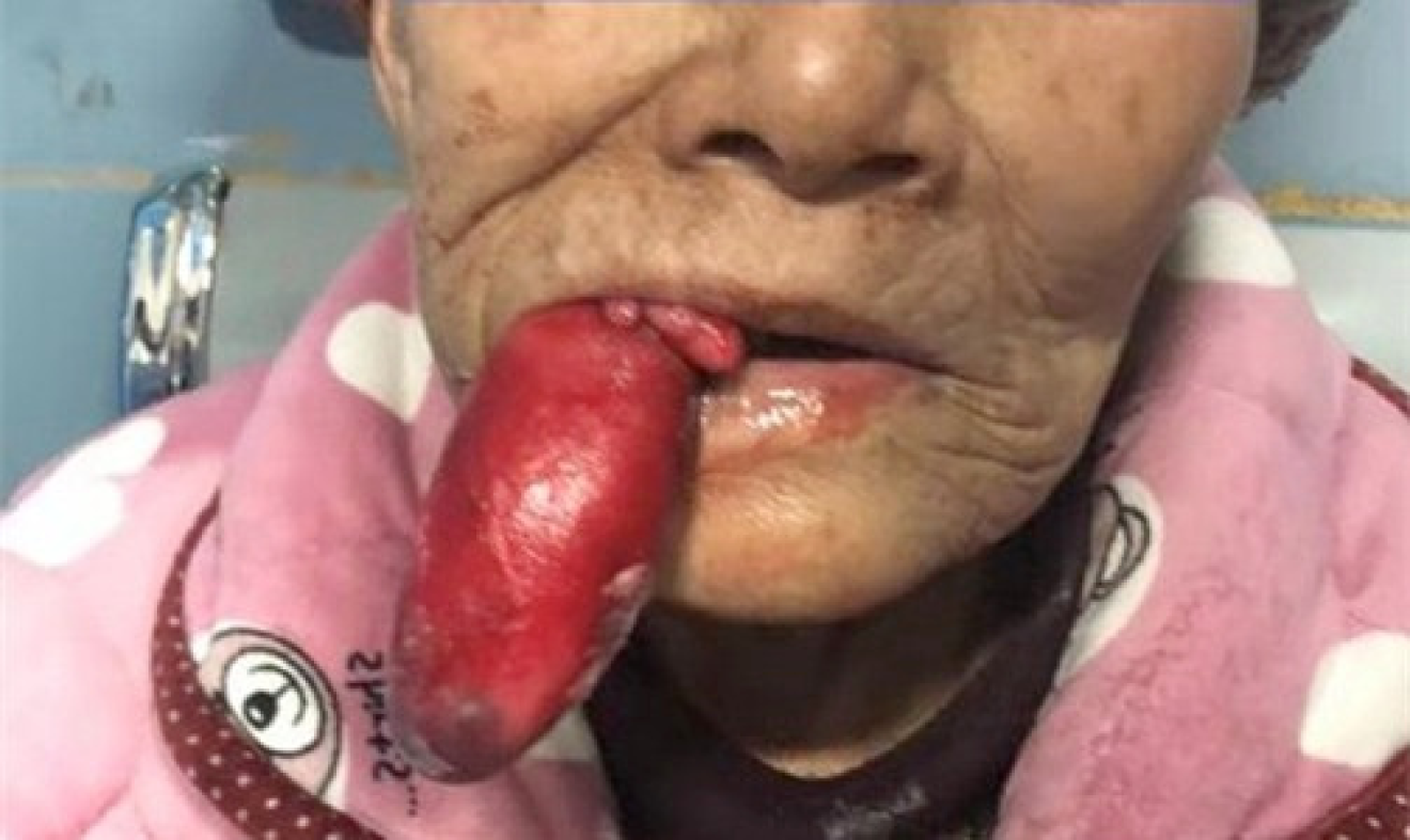
Figure 1 Mass protruding from the patient’s mouth.
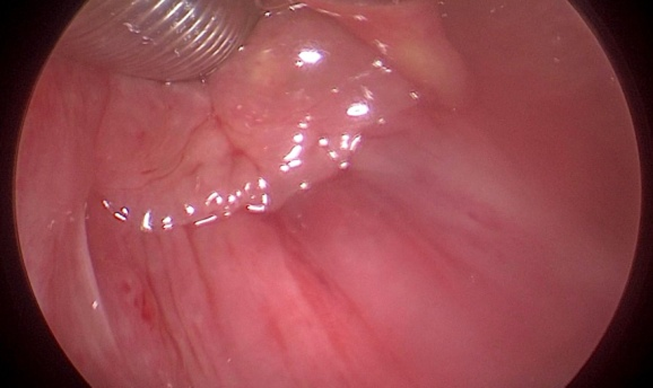
Figure 2 Root of lipoma was located in the posterior wall of the hypopharynx.
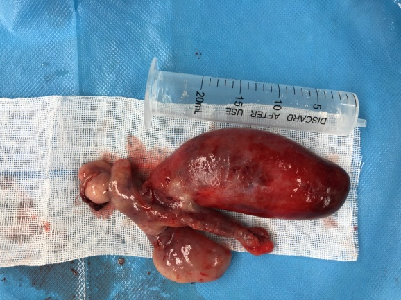
Figure 3 Lipoma in contrast with a 20 mL syringe.
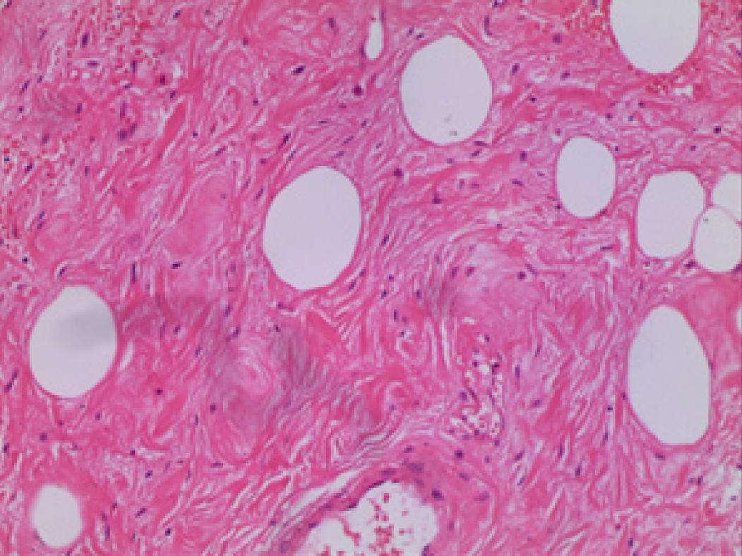
Figure 4 Histopathology revealed the distribution of adipocytes in dense fibrous tissue.
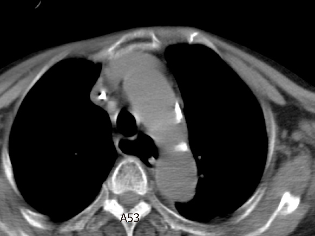
Figure 5 Postoperative chest computed tomography shows enlarged esophagus.
- Citation: Sun Q, Zhang CL, Liu ZH. An extremely rare pedunculated lipoma of the hypopharynx: A case report. World J Clin Cases 2019; 7(17): 2652-2657
- URL: https://www.wjgnet.com/2307-8960/full/v7/i17/2652.htm
- DOI: https://dx.doi.org/10.12998/wjcc.v7.i17.2652


