Copyright
©The Author(s) 2019.
World J Clin Cases. Aug 26, 2019; 7(16): 2401-2405
Published online Aug 26, 2019. doi: 10.12998/wjcc.v7.i16.2401
Published online Aug 26, 2019. doi: 10.12998/wjcc.v7.i16.2401
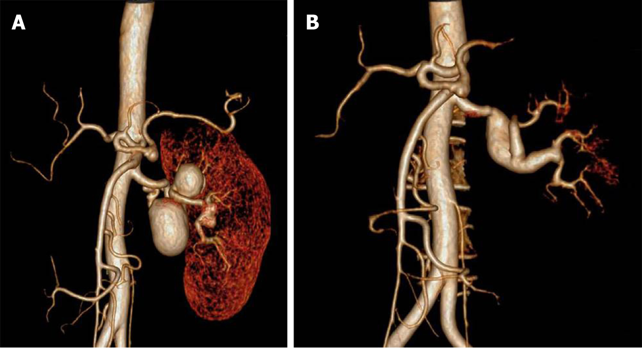
Figure 1 Computed tomography angiography.
A: Preoperative computed tomography angiography. Triple complicated multiple renal artery aneurysms with a maximum size of 4 cm, distal renal artery bifurcation, and branches were involved; B: Three-year follow-up computed tomography angiography. The saphenous vein graft was patent with aneurysmal degeneration.
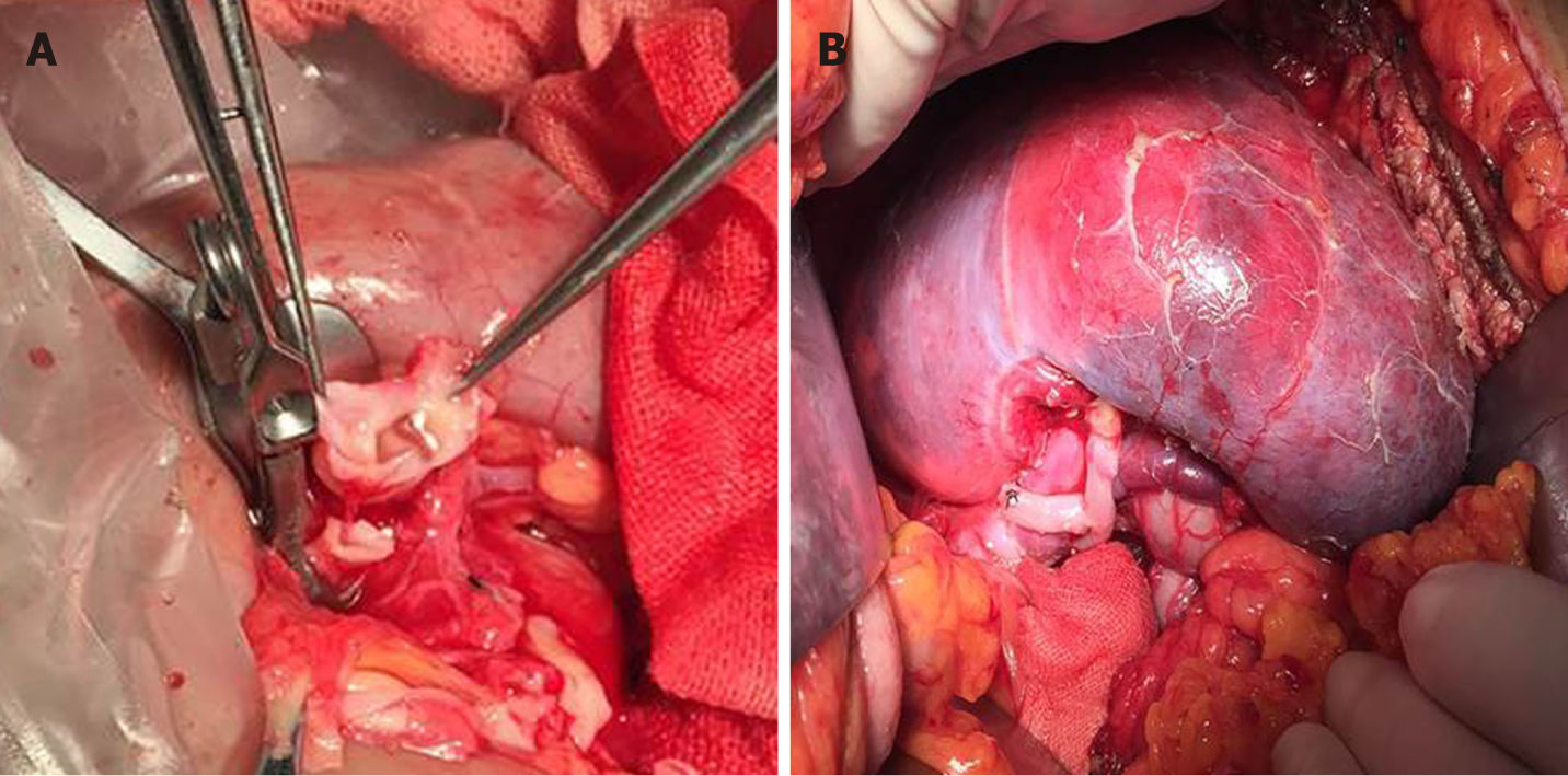
Figure 2 Surgical images.
A: Two residual distal branches were conjoined, creating a common patch; the distal anastomosis was performed end-to-end between the common patch and a reversed saphenous vein graft; B: Another branch originating from the second aneurysm was anastomosed end-to-side to the lateral wall of the saphenous vein graft.
- Citation: Chen XY, Zhao JC, Huang B, Yuan D, Yang Y. Ex vivo revascularization of renal artery aneurysms in a patient with solitary kidney: A case report. World J Clin Cases 2019; 7(16): 2401-2405
- URL: https://www.wjgnet.com/2307-8960/full/v7/i16/2401.htm
- DOI: https://dx.doi.org/10.12998/wjcc.v7.i16.2401


