Copyright
©The Author(s) 2019.
World J Clin Cases. Aug 26, 2019; 7(16): 2374-2383
Published online Aug 26, 2019. doi: 10.12998/wjcc.v7.i16.2374
Published online Aug 26, 2019. doi: 10.12998/wjcc.v7.i16.2374
Figure 1 Radiographic findings.
The computed tomography showed there were many enlarged lymph nodes in the chest, pulmonary atelectasis, and infection in the left lung. A: Transverse section; B: Coronal plane; C: Sagittal plane.
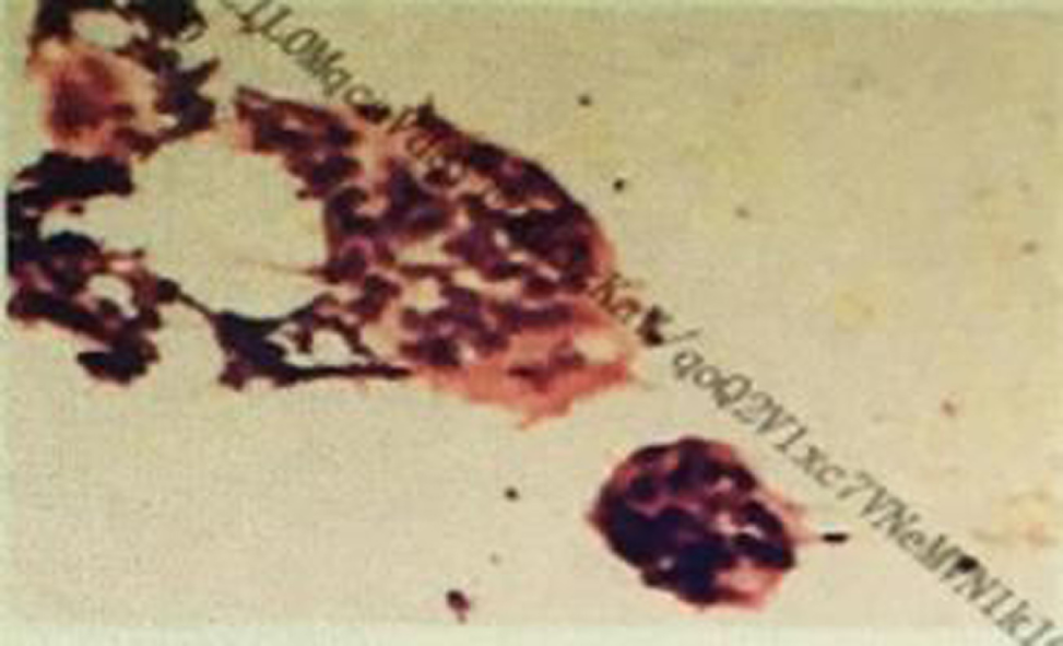
Figure 2 Biopsy of neck lymph node.
There are a small number of lymphoid cells and multinucleated giant cells and no malignant cells. Pathological diagnosis: (the left neck lymph node fine-needle aspiration smear). Considering the lymph node granulomatous lesions.
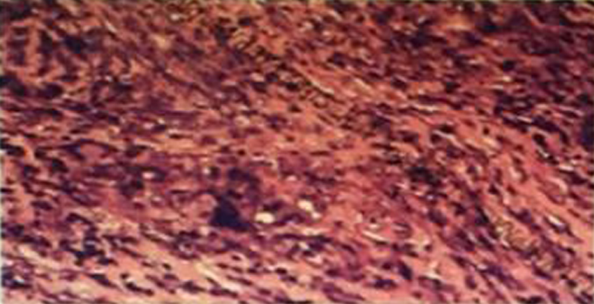
Figure 3 Secondary biopsy of supraclavicular lymph node.
Lymph nodes with widespread degeneration and necrosis, and there are many spores and small quantities of hyphae in these tissues. There are many giant cell granuloma in the peripheral lymphoid tissues. Pathological diagnosis: (the left supraclavicular lymph node fine-needle aspiration smear). The diagnosis conformed lymph nodes fungal disease.
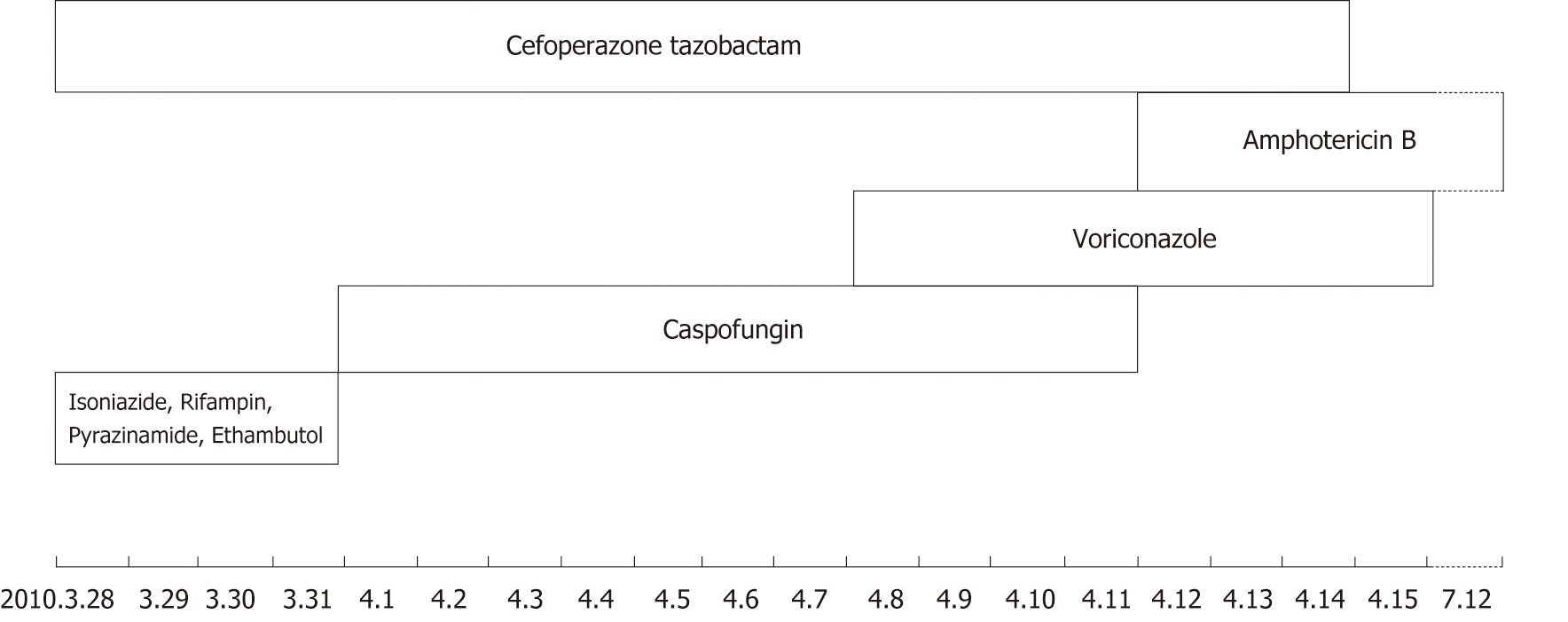
Figure 4 Timeline summarizing drug intervention.
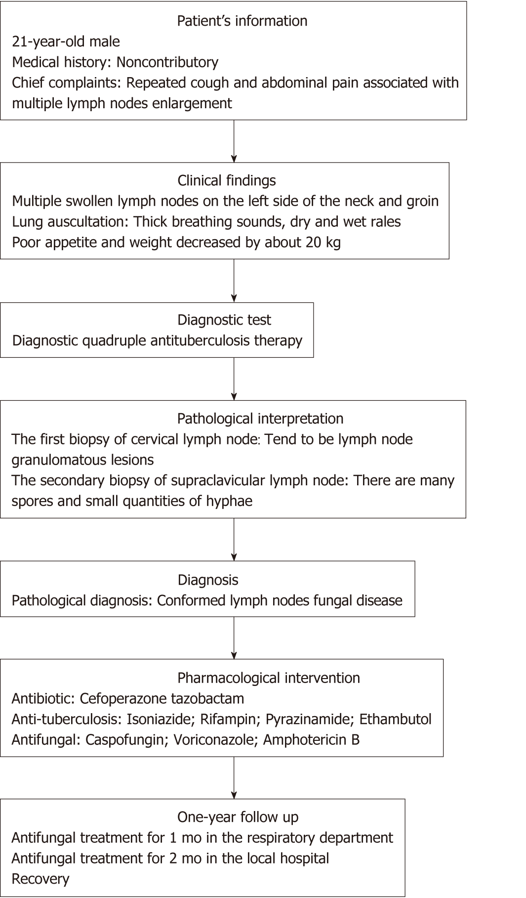
Figure 5 Timeline summarizing patient’s information, clinical findings, diagnostic tests, diagnosis, pharmacological intervention, and follow up.
- Citation: Xiao XF, Wu JX, Xu YC. Treatment of invasive fungal disease: A case report. World J Clin Cases 2019; 7(16): 2374-2383
- URL: https://www.wjgnet.com/2307-8960/full/v7/i16/2374.htm
- DOI: https://dx.doi.org/10.12998/wjcc.v7.i16.2374


