Copyright
©The Author(s) 2019.
World J Clin Cases. Aug 26, 2019; 7(16): 2352-2359
Published online Aug 26, 2019. doi: 10.12998/wjcc.v7.i16.2352
Published online Aug 26, 2019. doi: 10.12998/wjcc.v7.i16.2352
Figure 1 Computed tomography scans before treatment.
A: Non-contrast abdominal computed tomography (CT) showed a 6.4 cm × 6.0 cm, nearly circular, heterogeneous lesion, owning indistinct borders, located in the head of the pancreas. B: Contrast-enhanced CT imaging indicated that the fat containing tumor (-109 ± 19.2 HU in the tumor and 47.9 ± 14.9 HU in the liver) had a few fibroreticular septa within it, and the surrounding parenchyma of the mass could be slightly enhanced (arrow).
Figure 2 Magnetic resonance scans before treatment.
A and B: Both fat-suppressed T1-weighted and fat-suppressed T2-weighted images showed a loss in signal intensity (arrow), which indicated the mass mainly composed of adipose tissue. And a few fibroreticular septa could be seen within the lesion. The boundary between the lesion and the pancreas was unclear.
Figure 3 Coronary magnetic resonance scan and magnetic resonance cholangiopancreatography before treatment.
A: Magnetic resonance imaging showed a fat-signal lobulated tumor compressing her duodenum (arrow); B: Magnetic resonance cholangiopancreatography demonstrated that there was no dilatation and stenosis in the intrahepatic bile duct and the pancreatic duct, but the middle and lower part of the common bile duct was partially compressed (arrow).
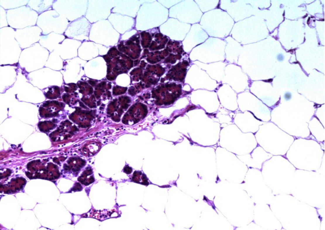
Figure 4 Final pathological examination.
Mature adipocytes were noted adjacent to the pancreatic parenchyma (original magnification, × 100).
- Citation: Xiao RY, Yao X, Wang WL. A huge pancreatic lipoma mimicking a well-differentiated liposarcoma: A case report and systematic literature review. World J Clin Cases 2019; 7(16): 2352-2359
- URL: https://www.wjgnet.com/2307-8960/full/v7/i16/2352.htm
- DOI: https://dx.doi.org/10.12998/wjcc.v7.i16.2352


