Copyright
©The Author(s) 2019.
World J Clin Cases. Aug 26, 2019; 7(16): 2346-2351
Published online Aug 26, 2019. doi: 10.12998/wjcc.v7.i16.2346
Published online Aug 26, 2019. doi: 10.12998/wjcc.v7.i16.2346
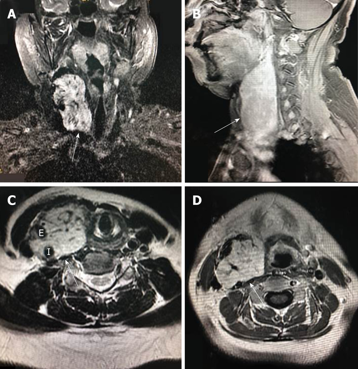
Figure 1 Enhanced magnetic resonance imaging.
A: Coronal section (arrow indicates enhancement); B: Sagittal section (arrow); C, D: Axial section showing blood flow in the tumor (arrows). E: External carotid artery; I: Internal carotid artery.
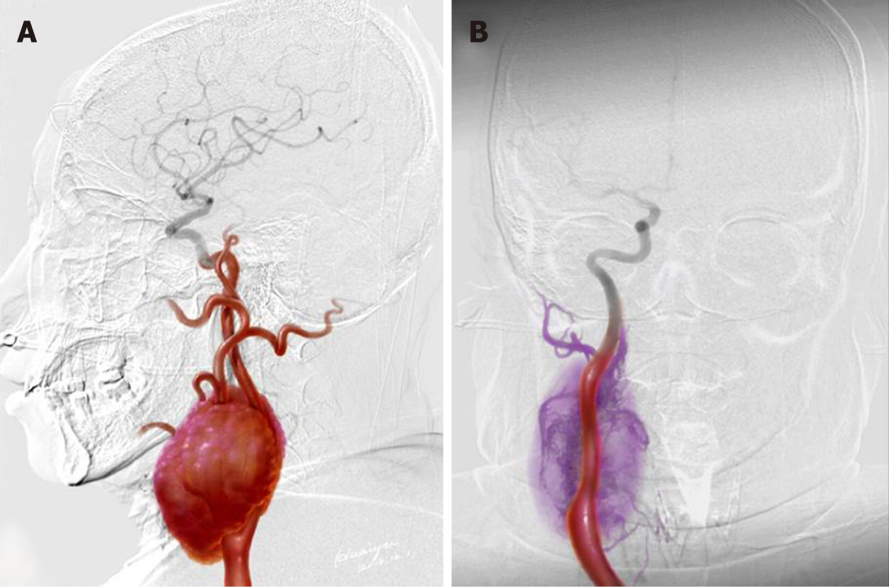
Figure 2 Hypervascular carotid body tumor spans and remove of tumor.
A: The hypervascular carotid body tumor spans from C1 to C7, and adheres to the right common carotid artery, right internal carotid artery and right external carotid artery; B: Purple: Removed tumor and external carotid artery segment; Red: Right common carotid artery and right internal carotid artery.
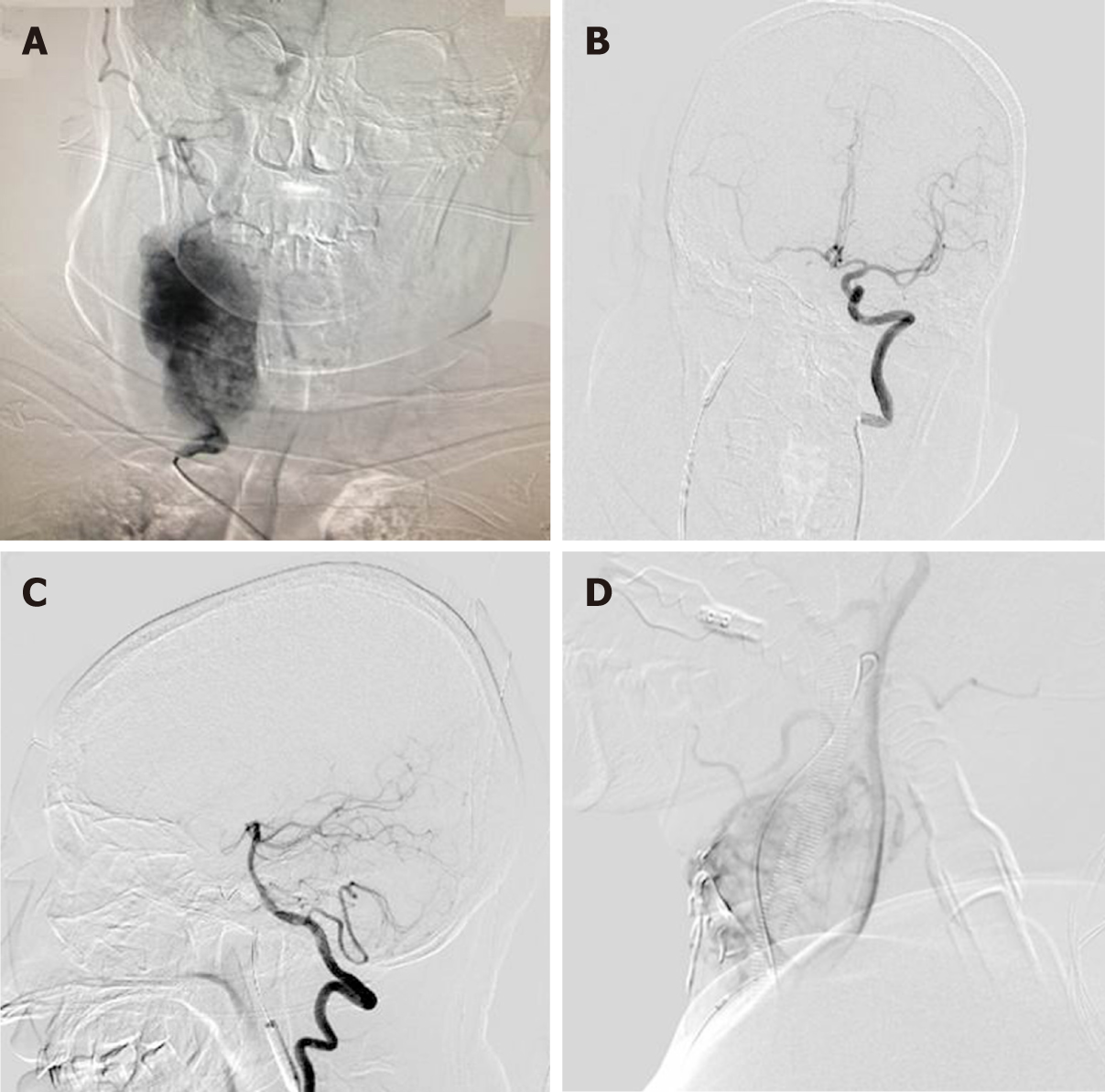
Figure 3 Preoperative angiography and balloon test occlusion.
A: Right carotid artery angiography revealed a hypervascular tumor; B: Balloon test occlusion showed that the anterior communicating artery was open and the right internal carotid artery blood supply was compensated; C: The left vertebral artery angiography showed the posterior communicating artery was open; D: Embolization of the small feeding artery of carotid body tumor.
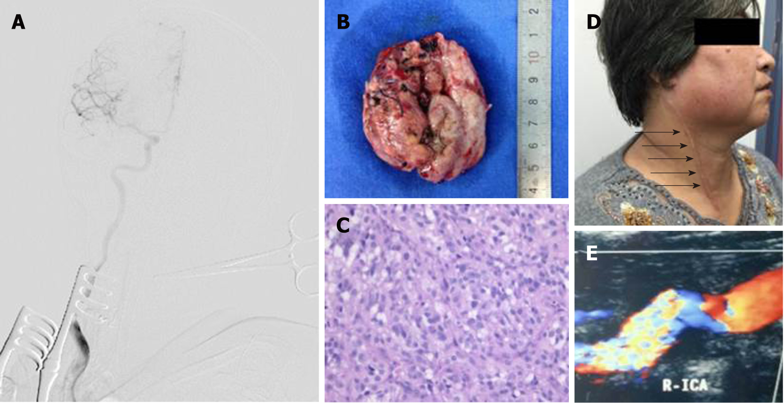
Figure 4 The tumor was completely removed and the right internal carotid artery flow was unobstructed.
A: Intraoperative angiography showed that the internal carotid artery was unobstructed after the carotid body tumor was completely removed; B: The tumor volume decreased significantly due to blockade of blood supply; C: HE staining showed paraganglioma (× 400); D: Ultrasonography at 2 wk after the surgery (arrows: The site of surgical incision); EL 4-year follow-up.
- Citation: Li MQ, Zhao Y, Sun HY, Yang XY. Large carotid body tumor successfully resected in hybrid operating theatre: A case report. World J Clin Cases 2019; 7(16): 2346-2351
- URL: https://www.wjgnet.com/2307-8960/full/v7/i16/2346.htm
- DOI: https://dx.doi.org/10.12998/wjcc.v7.i16.2346


