Copyright
©The Author(s) 2019.
World J Clin Cases. Aug 26, 2019; 7(16): 2336-2340
Published online Aug 26, 2019. doi: 10.12998/wjcc.v7.i16.2336
Published online Aug 26, 2019. doi: 10.12998/wjcc.v7.i16.2336
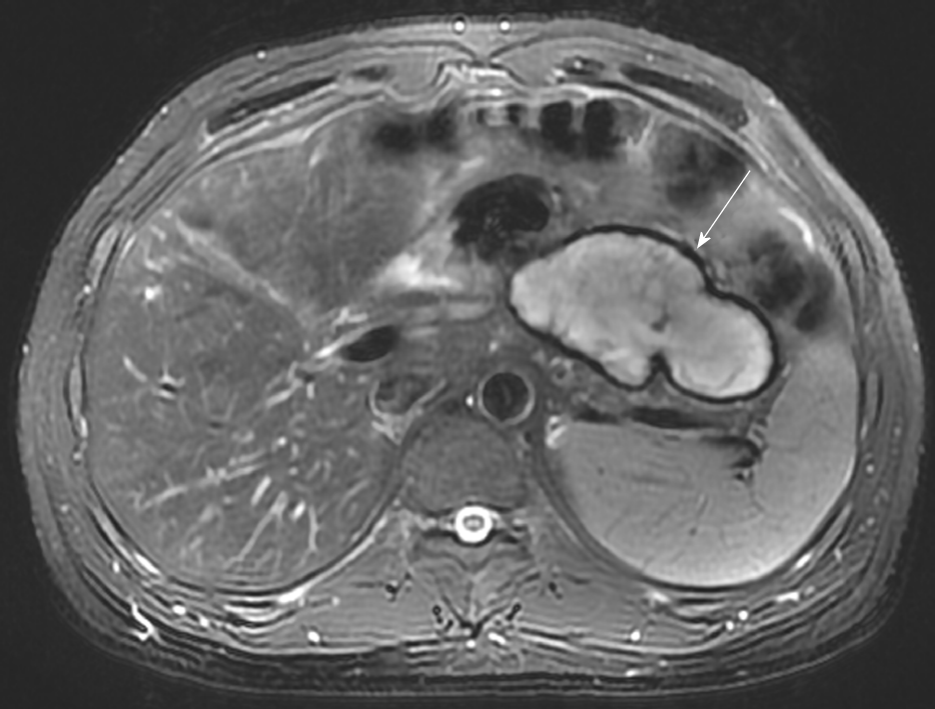
Figure 1 An encapsulated effusion was found in the abdominal cavity of the patient in Case 1 at the 3-mo follow-up.
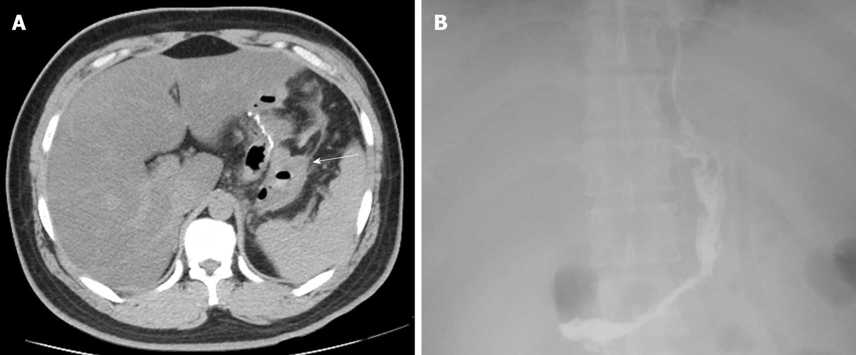
Figure 2 Imaging findings in Case 2.
A: A blood clot and peritoneal cavity free air were found; B: No gastric leakage was detected by upper gastrointestinal radiography.
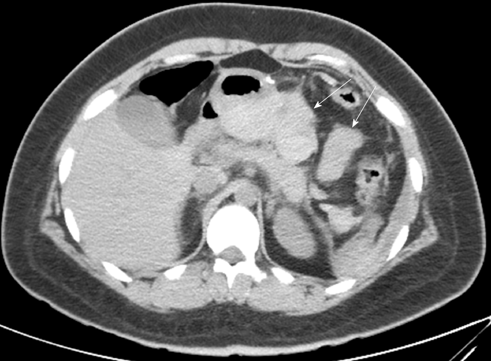
Figure 3 Blood clots behind the stomach were found in Case 3.
- Citation: Liu Y, Li MY, Zhang ZT. Role of abdominal drainage in bariatric surgery: Report of six cases. World J Clin Cases 2019; 7(16): 2336-2340
- URL: https://www.wjgnet.com/2307-8960/full/v7/i16/2336.htm
- DOI: https://dx.doi.org/10.12998/wjcc.v7.i16.2336


