Copyright
©The Author(s) 2019.
World J Clin Cases. Aug 26, 2019; 7(16): 2302-2308
Published online Aug 26, 2019. doi: 10.12998/wjcc.v7.i16.2302
Published online Aug 26, 2019. doi: 10.12998/wjcc.v7.i16.2302
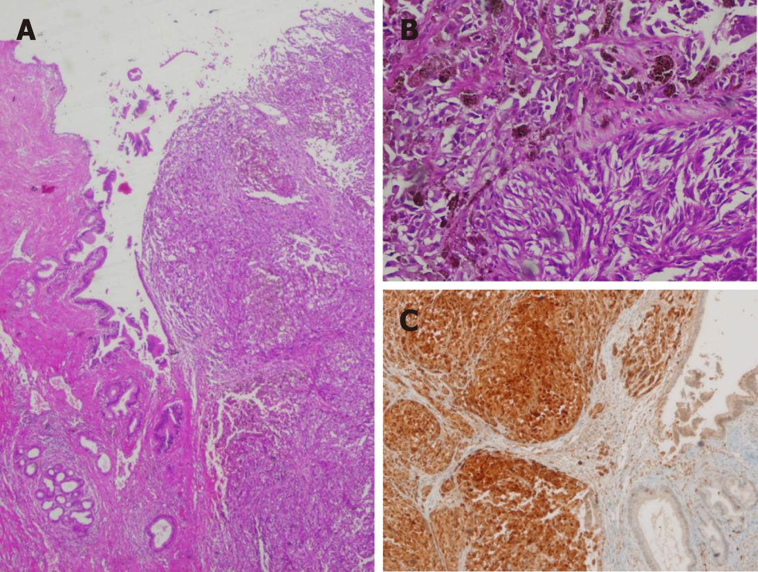
Figure 1 Primary melanoma of the biliary tract.
A: The melanoma formed a polypoid mass that infiltrates the wall of the extrahepatic bile duct; B: At high magnification, some malignant melanocytes revealed finely granular cytoplasmic melanin; C: Tumour cells revealed strong immunopositivity for the HMB45 antigen.
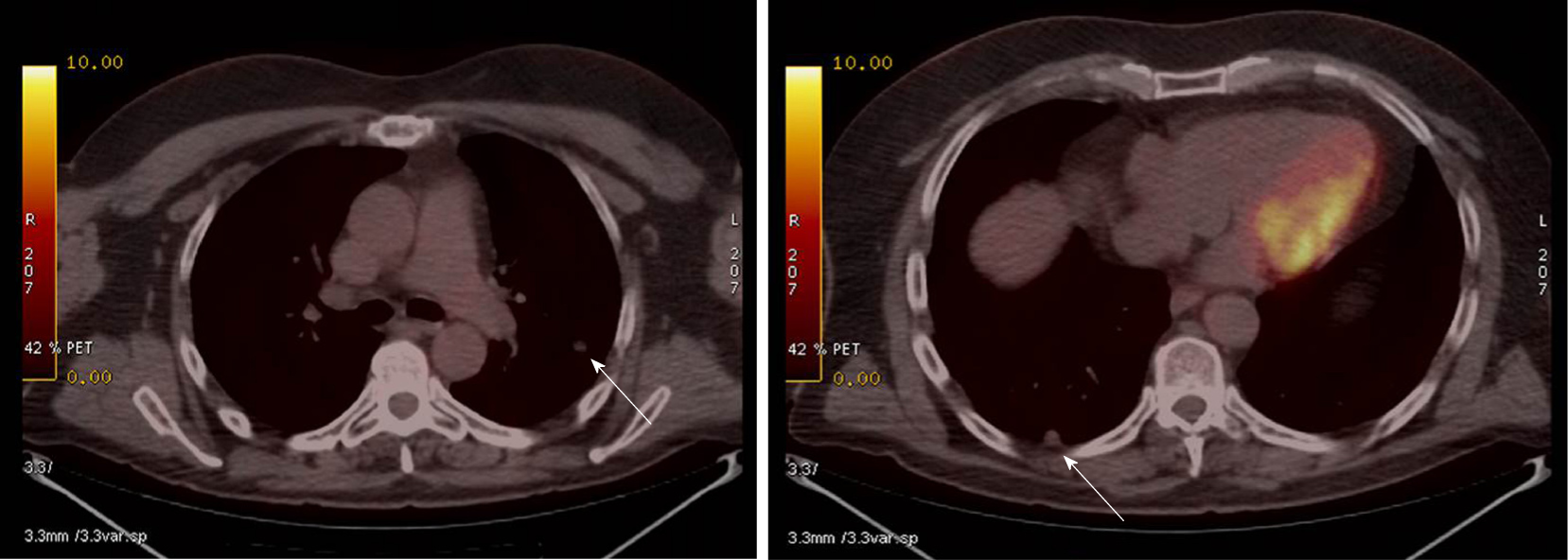
Figure 2 Thoracic positron emission tomography/computed tomography scan showing metastatic nodules (arrows).
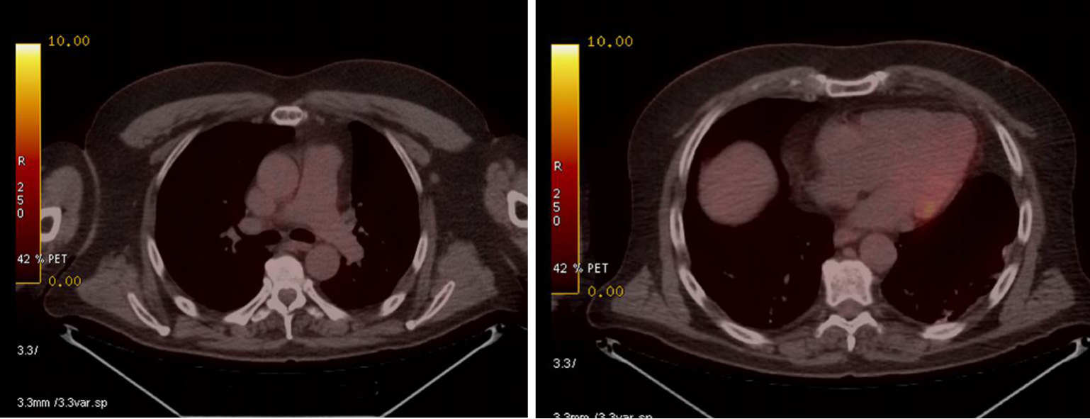
Figure 3 Thoracic positron emission tomography/computed tomography scan with no evidence of metastases.
- Citation: Cameselle-García S, Pérez JLF, Areses MC, Castro JDF, Mosquera-Reboredo J, García-Mata J. Primary malignant melanoma of the biliary tract: A case report and literature review. World J Clin Cases 2019; 7(16): 2302-2308
- URL: https://www.wjgnet.com/2307-8960/full/v7/i16/2302.htm
- DOI: https://dx.doi.org/10.12998/wjcc.v7.i16.2302


